Capsule endoscope having a self-cleaning surface and method of using the same

a self-cleaning, endoscope technology, applied in the field of endoscopes, can solve the problem that the images generated by the endoscopes do not have much useful information, and achieve the effect of easy and successful cleaning

Active Publication Date: 2016-10-06
ANKON MEDICAL TECH (SHANGHAI) CO LTD
View PDF5 Cites 27 Cited by
  • Summary
  • Abstract
  • Description
  • Claims
  • Application Information

AI Technical Summary

Benefits of technology

The present invention is about a capsule endoscope that can prevent itself from getting covered with residue or mucous in a patient's digestive tract. It has a special surface that doesn't stick to anything. Even if there is a little bit of residue or mucous in the patient's digestive tract, the special surface makes it easy to clean the capsule before taking pictures.

Problems solved by technology

If accidentally, the residue landed on the surface of an imaging device or a lighting unit is accidentally covered, then the images generated by capsule endoscope will not have much useful information.

Method used


Image

  • Capsule endoscope having a self-cleaning surface and method of using the same
  • Capsule endoscope having a self-cleaning surface and method of using the same
  • Capsule endoscope having a self-cleaning surface and method of using the same

Examples


example 1

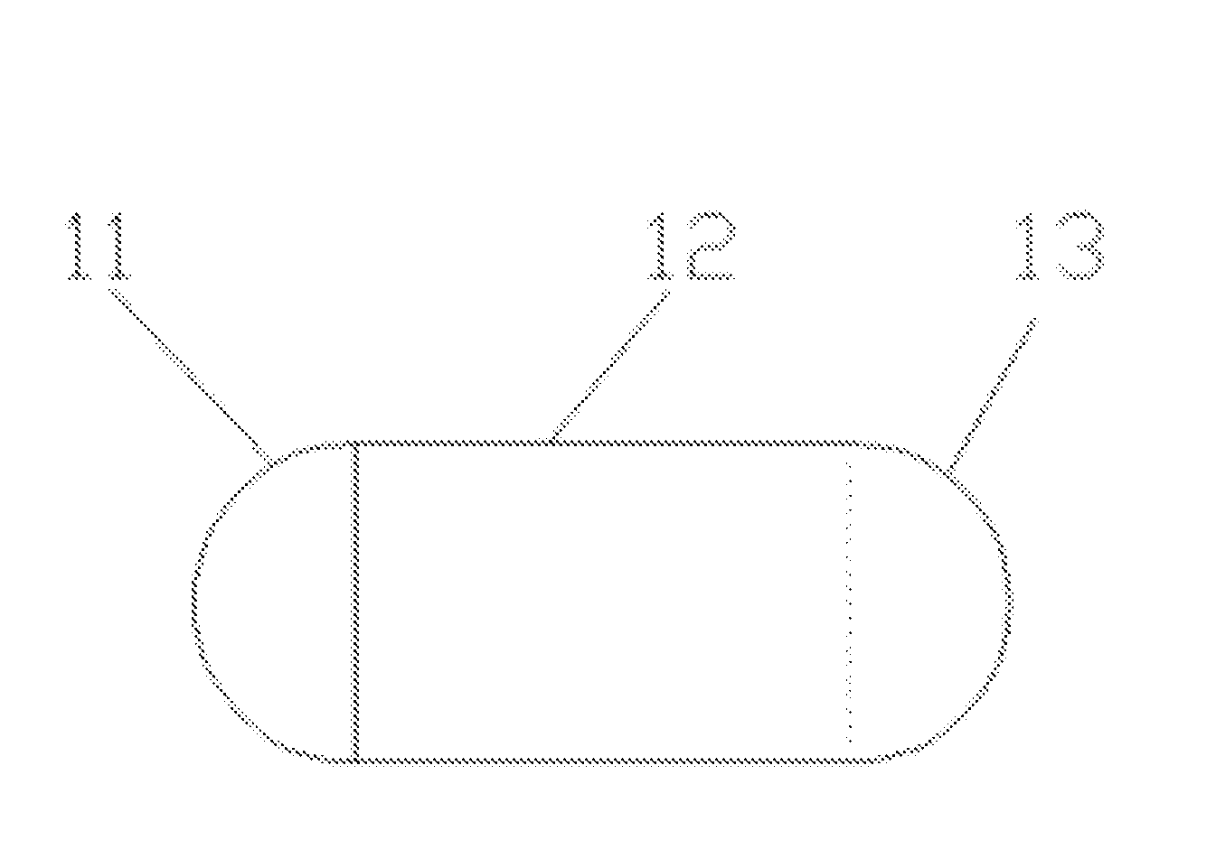
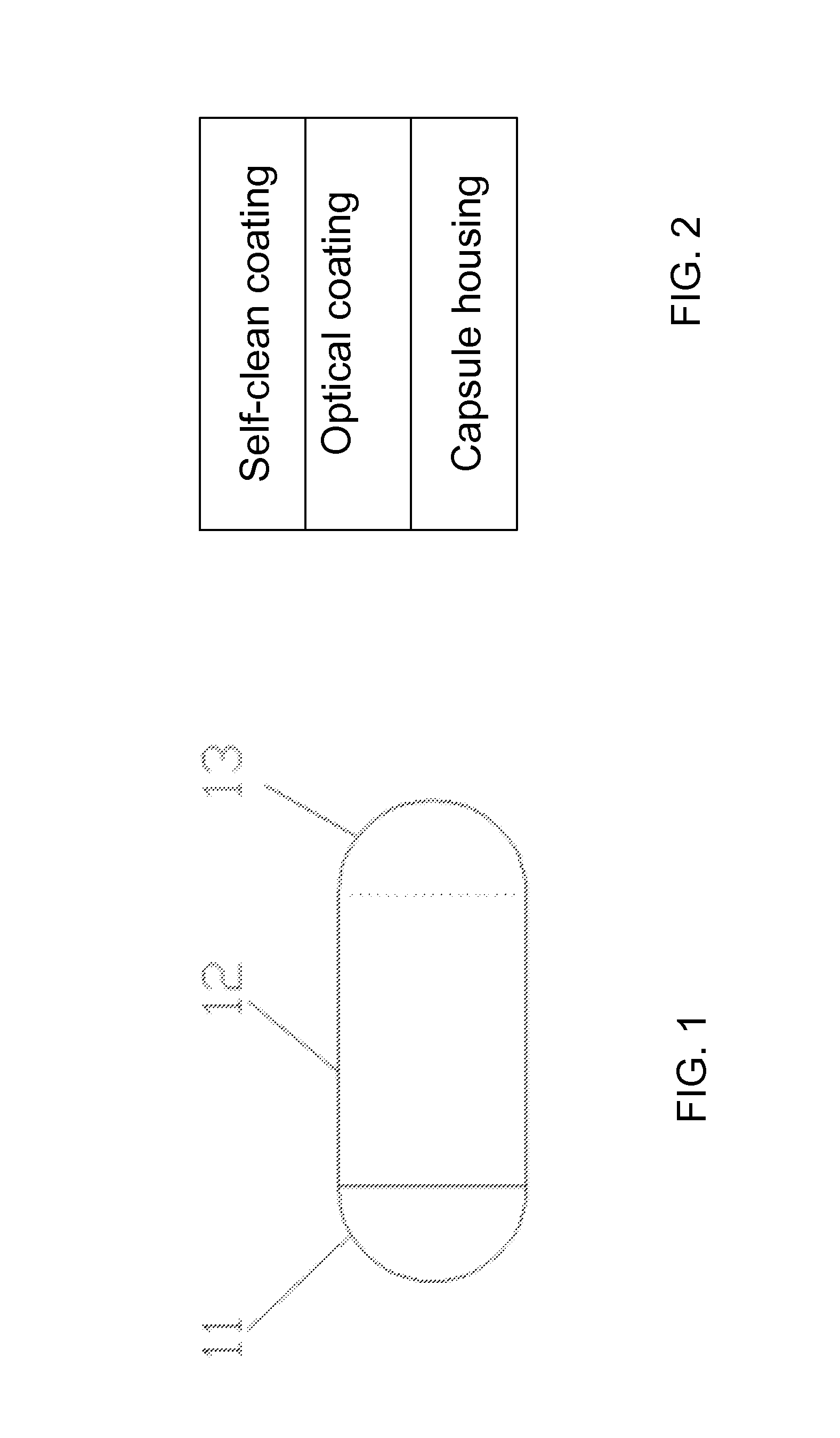
[0056]A capsule endoscope, having a housing including a front end 11, capsule body 12 and rear end 13, is provided. The front end is transparent. The imaging device is placed inside the housing underneath the surface of front end. The front end 11, capsule body 12 and rear end 13 are all made polycarbonate and have surface modification including a layer of self-clean coating. The exterior surface of the front end 11 is illustrated as FIG. 4. The surface of the front end 11 is sequentially coated with TiO2, SiO2, TiO2, SiO2, TiO2, and SiO2. On top of the last SiO2, a layer of nano scale CF3(CF2)5(CH2)2Si— is grown. The resulted self-clean surface is a low surface energy surface having a contact angle more than 130°. The self-clean surface can effectively prevent residues and mucus accumulated on capsule surface and improve the image quality. The layer of CF3(CF2)5(CH2)2Si— is formed on the capsule surface either in gas phase or in liquid phase. The resulted surface covered by the flu...

example 2

[0057]A capsule endoscope, having a housing including a front end 11, capsule body 12 and rear end 13, is provided. The front end is transparent. The imaging device is placed inside the housing underneath the surface of front end. The front end 11, capsule body 12 and rear end 13 are all made polycarbonate and only the front end 11 and capsule body have are coated with a layer of self-clean coating. The rear end is not covered by the self-clean coating. The self-clean coating is formed by reacting CF3(CF2)5(CH2)2SiCl3 with a hetero atom of the surface of the housing. This differential surface treatment leads a capsule endoscope, whose rear end having a contact angle smaller than the contact angle of the front end and capsule body. Such capsules can be navigated to a target area having a liquid, to clean the front end thereof by swirling the front end in the liquid or rubbing the front end against the walls of the target area with or without a liquid.

example 3

[0058]A capsule endoscope, having a housing including a front end 11, capsule body 12 and rear end 13, is provided. The housing is made of PSF. The entire housing including front end 11, capsule body 12 and rear end 13 are all coated with optical coatings including TiO2, SiO2, TiO2, SiO2, TiO2, and SiO2. On top of the last SiO2, a layer of nano scale CF3(CF2)5(CH2)2Si— is grown. This surface treatment leads to a capsule endoscope which is not easy to collect any residue or mucous in the digestive track.


PUM


Abstract

The present invention discloses a capsule endoscope and a method to prevent the capsule from being covered by a residue or mucous in a patient's digestive tract. The capsule endoscope comprises an improved surface, preventing a residue or mucous to adhere onto the surface of the capsule, and in the unlikely event that a residue or a mucous is collected by the capsule the improved surface can allow an easy and successful clean before an image is taken.

Description

CROSS REFERENCE TO RELATED APPLICATIONS[0001]This application is a provisional application.TECHNICAL FIELD[0002]The invention relates to medical apparatus and instruments, specifically refers to a capsule endoscope having a surface modification for self-cleaning.BACKGROUND OF THE INVENTION[0003]Capsule endoscope is a miniaturized camera placed inside a capsule-shaped housing. When a patient swallows such a camera pill, the capsule endoscope travels along the patient's gastrointestinal tract and takes a series of pictures of the interior of the patient gastrointestinal tract. While the capsule endoscope passing through the patient's digestive system, the images taken would be simultaneously transmitted outside of the patient's body to a receiver, and then doctors use the image data for real-time medical examinations.[0004]Capsule endoscopy not only has shown clear advantage in examining a patient's small intestine, where the areas or portions of the gastrointestinal tract are not rea...

Claims


Application Information

Patent Timeline
Patent Type & Authority Applications(United States)
IPC IPC(8): A61B1/00A61B1/06A61B1/12A61B1/04
CPCA61B1/00158A61B1/126A61B1/0661A61B1/041
Inventor YE, QINGDUAN, XIAODONG
Owner ANKON MEDICAL TECH (SHANGHAI) CO LTD
Why Eureka
Try Eureka