Beamforming techniques for ultrasound microcalcification detection

Active Publication Date: 2016-10-13
KONINKLJIJKE PHILIPS NV
View PDF14 Cites 1 Cited by
  • Summary
  • Abstract
  • Description
  • Claims
  • Application Information

AI Technical Summary

Benefits of technology

[0009]Channel data contain much more information than B-mode images obtained after ultrasound receive beamforming. Therefore, channel-data-based beamforming techniques can provide better sensitivity and/or specificity for microcalcification detection than image processing techniques. The novel technology proposed herein enhances the contrast between microcalcifications and the background, i.e., body tissue in which the calcifications reside, typically s

Problems solved by technology

However, mammography shows poor performance in dense breast tissue, the non-fatty portion of the breast.
Also, despite measures to reduce the dose, mammography exposes the patient to ionizing radiation.
In addition, there i

Method used


Image

  • Beamforming techniques for ultrasound microcalcification detection
  • Beamforming techniques for ultrasound microcalcification detection
  • Beamforming techniques for ultrasound microcalcification detection

Examples


Embodiment Construction

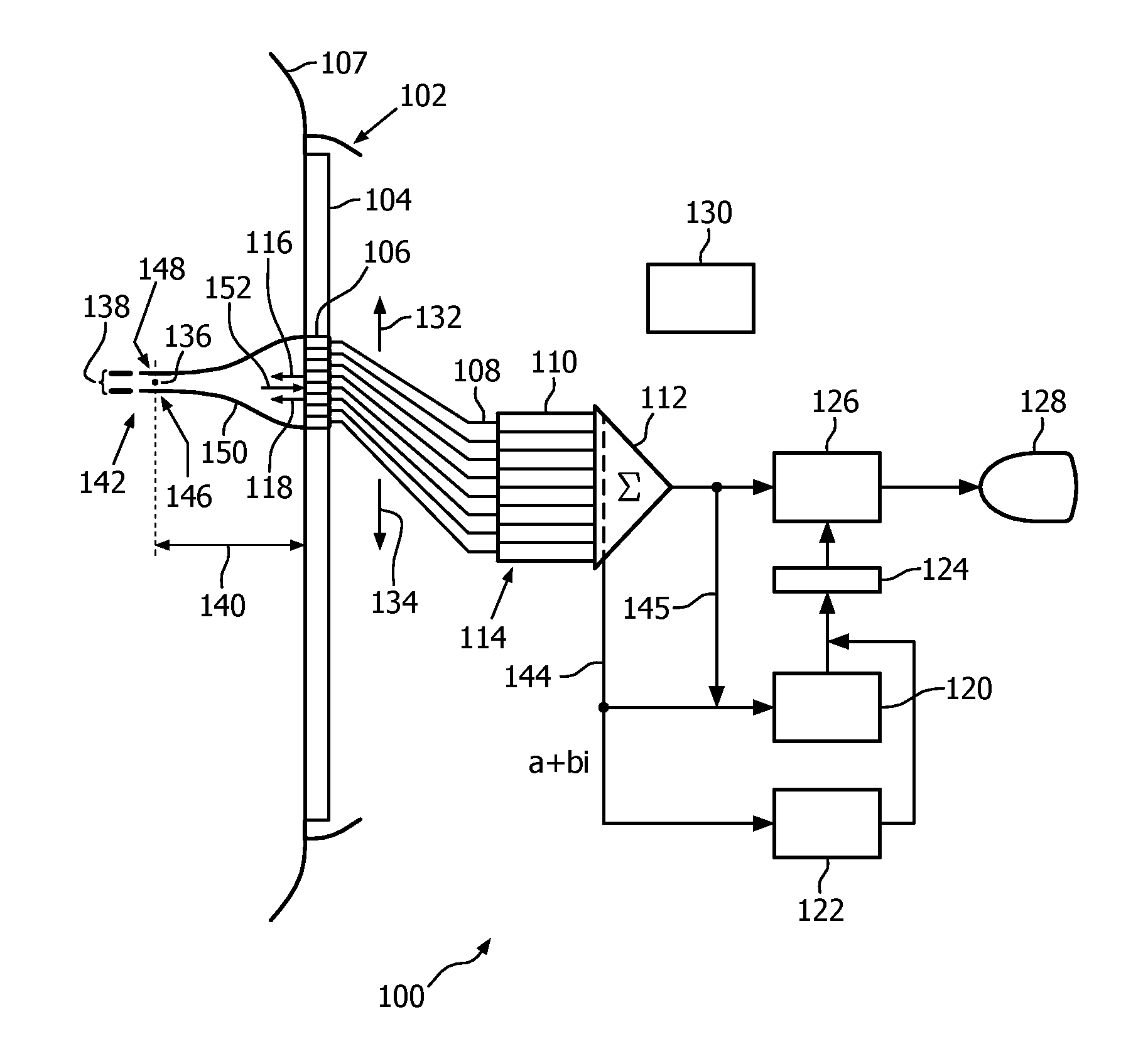
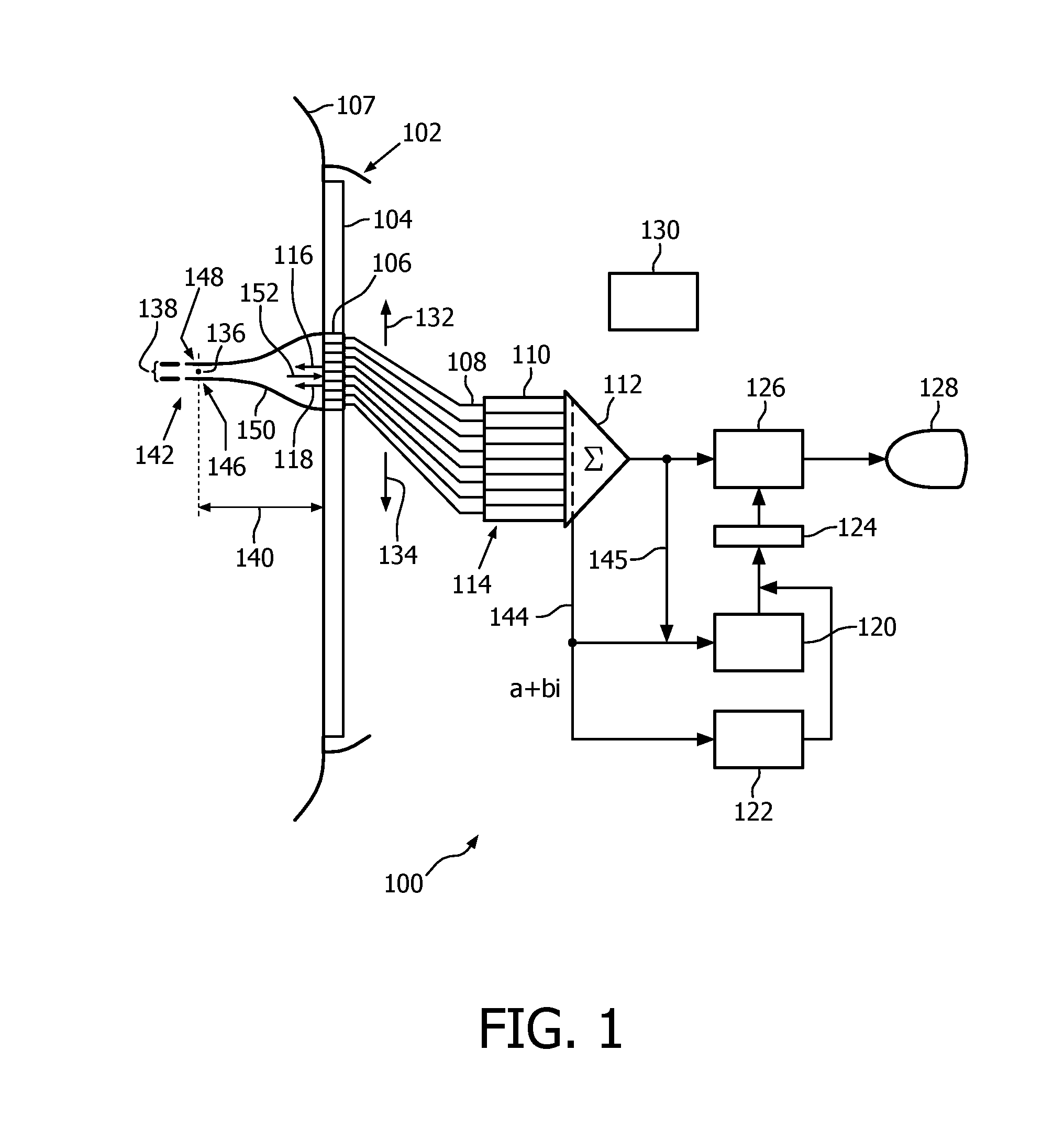
[0017]FIG. 1 depicts, by way of illustrative and non-limitative example, a medical ultrasound acquisition-data analysis device characterizable as an ultrasonic microcalcification identification device 100.

[0018]The device 100 includes an ultrasound imaging probe 102 having an array 104 of transducer elements 106. The probe 102 is shown as pressed into acoustic contact with breast tissue 107.

[0019]The device 100 further includes channels 108 in connection with the elements 106. The channels have respective sample delay elements 110. The latter connect to a coherent summer 112, the summer and the receive delay elements 110 together constituting a receive beamformer 114. The delay elements 110 may be augmented to also provide amplitude weighting. Delay elements for steering and focusing transmit beams 116, 118 can be implemented and are not shown. The multiple transmit beams 116, 118 may be parallel, or may be differently angled, i.e., steered.

[0020]Also included in the device 100 are ...


PUM


Abstract

A medical ultrasound acquisition-data analysis device acquires channel data (144) via ultrasound received on the channels, uses the acquired channel data to estimate data coherence and derive dominance of an eigen-value of a channel covariance matrix and, based on the estimate and dominance, distinguishes microcalcifications (142) from background. Microcalcifications may then be made distinguishable visually on screen via highlighting, coloring, annotation, etc. The channel data operable upon by the estimating may have been subject to beamforming delays and may be summed in a beamforming procedure executed in the estimating. In the estimating and deriving, both field point-by-field point, multiple serial transmits (116, 118) may be used for each field point. In one embodiment results of the estimating and deriving are multiplied point-by-point and submitted to thresholding.

Description

CROSS-REFERENCE TO PRIOR APPLICATION[0001]This application claims the benefit of U.S. Provisional Patent Application No. 61 / 803,634, filed Mar. 20, 2013 and U.S. Provisional Patent Application No. 61 / 907,022, filed Nov. 21, 2013. These applications are hereby incorporated by reference herein.FIELD OF THE INVENTION[0002]The present invention relates to medical imaging for identifying microcalcifications and, more particularly, to use of ultrasound as the imaging modality.BACKGROUND OF THE INVENTION[0003]Mammography's success for screening for breast cancer is mostly attributed to its capability for reliably imaging calcifications, an important marker for early stage breast cancer. Microcalcifications are present in 60-80% of breast cancers and are a reliable indicator of both benign and malignant lesions. The key diagnostic features of microcalcifications are their location, number, size, morphology, distribution, pattern, and relation to a mass. These features help stratify the risk...

Claims


Application Information

Patent Timeline
IPC IPC(8): A61B8/08A61B8/00
CPCA61B8/085A61B8/461A61B8/5207A61B8/0825A61B8/5223G16H50/30
Inventor ROBERT, JEAN-LUCHUANG, SHENG-WENRADULESCU, EMIL GEORGEVIGNON, FRANCOIS GUY GERARD MARIEERKAMP, RAMON QUIDO
Owner KONINKLJIJKE PHILIPS NV
Why Eureka
Try Eureka