
Systems and methods for extracting prognostic image features

Active Publication Date: 2017-01-19
DUKE UNIV
View PDF0 Cites 11 Cited by
  • Summary
  • Abstract
  • Description
  • Claims
  • Application Information

AI Technical Summary

Benefits of technology

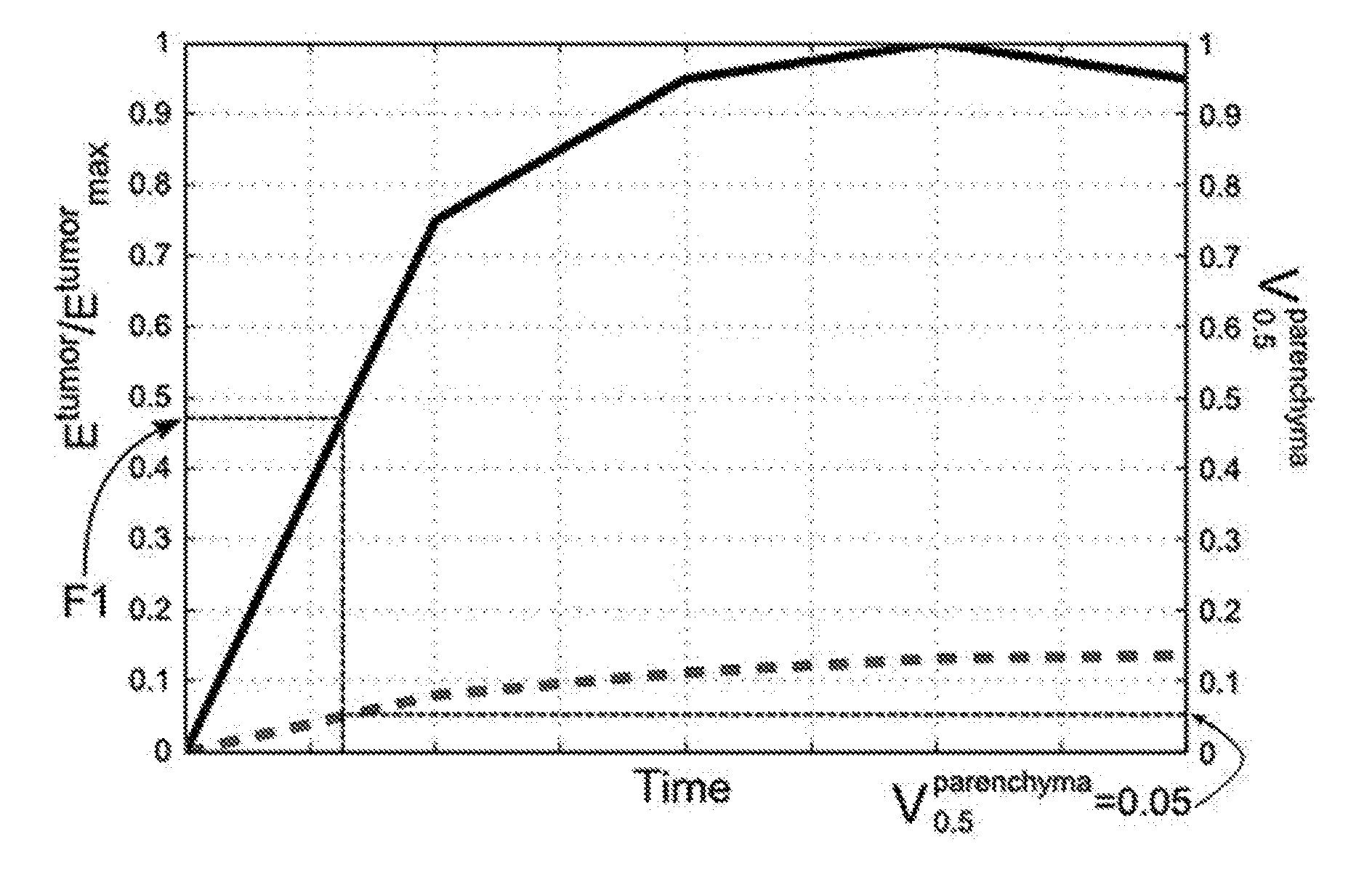
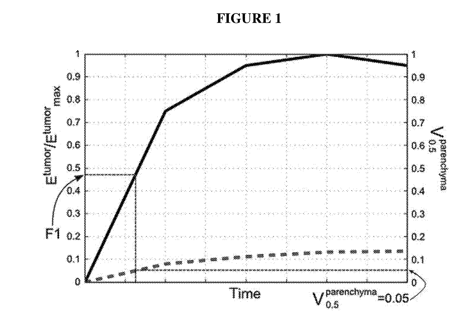
The patent text describes a method for predicting the risk of breast cancer progression using a marker called T11. The researchers found that a higher value of feature F1 was associated with an increased risk of breast cancer disease progression. These findings may help improve the diagnosis and treatment of breast cancer.

Problems solved by technology

The luminal B subtype has been shown to have significantly worse relapse-free survival rates than the luminal A subtype, with the increased risk limited to the early period after surgery.
Although differentiation of breast cancer into molecular subtypes is a potentially powerful tool to enable clinicians to better guide therapeutic interventions, genetic analysis presents a high barrier because of the cost, specialized equipment, and technical expertise needed to process each sample.
As a result, formal molecular subtype analysis is currently neither practical nor cost efficient for all patients with breast cancer.
Breast cancer subtype analysis is important for establishing initial treatment regimens, but clinicians must also make complicated decisions regarding how, when, and if to utilize chemotherapy, endocrine therapy, radiation therapy, and / or surgery.

Method used


Image

  • Systems and methods for extracting prognostic image features
  • Systems and methods for extracting prognostic image features
  • Systems and methods for extracting prognostic image features

Examples


example 1

Luminal B Breast Tumor Molecular Subtype is Associated with MRI Tumor Enhancement Dynamics

I. Study Methods

Patient Population

[0076]This was an institutional review board approval exempt study of information from a publically available database. We used the Breast Cancer Risk Assessment dataset recently made available by the Cancer Imaging Archive. The Cancer Imaging Archive dataset contains imaging studies for a subset of patients from the Cancer Genome Atlas. The Cancer Genome Atlas database contains full genomic sequencing and clinical data for the patients. The two databases were correlated by using unique patient identifiers.

[0077]A fellowship-trained breast imager (S.C.Y., with 9 years of experience reading breast MR images) marked the MR images by drawing a box containing the abnormality. The reader used a graphical user interface developed in our laboratory. The lesions were classified into four genomic subtypes as luminal A, luminal B, HER2-enriched, or basal-like. The subtyp...

example 2

Luminal A and Luminal B Molecular Subtypes are Associated with Imaging Features Extracted Following Routine Breast MRI

I. Study Methods

Patient Population

[0087]Institutional Review Board approval was secured for the collection and analysis of this Health Information Portability and Accountability Act compliant study with a waiver of informed consent. We retrospectively collected data from a total of 400 consecutive preoperative breast MRIs that were performed between September 2007 and June 2009. We excluded patients undergoing breast cancer treatment at the time of the MRI (e.g., chemotherapy or radiation therapy, N 29), and those with a history of elective breast surgery (e.g., implants or mammoplasty, N 19) or remote prior breast cancer (i.e., those having undergone previous definitive therapy, N 19). Patients without complete pathology data available for review (e.g., hormone receptor status) were excluded (N 42). There were subsequently 291 routine preoperative breast MRIs availa...

example 3

Breast Cancer Progression-Free Survival is Associated with MRI Tumor Enhancement Dynamics

I. Study Methods

Patient Population

[0102]Institutional Review Board (IRB) approval was secured for this study. We retrospectively collected data from 400 consecutive preoperative contrast enhanced breast MRIs from September 2007 through June 2009. Patients with a history of elective breast surgery (e.g., implants or mammoplasty, n=19) or remote prior breast cancer (i.e., those having undergone previous definitive therapy, n=19), as well as those undergoing breast cancer treatment at the time of the MRI (e.g., chemotherapy or radiation therapy, n=29) were excluded. Additionally, patients with missing pathology data (e.g., tumor grade) were excluded (n=42). This resulted in 291 patients with preoperative breast MRIs available for analysis. After a review of the imaging sequences, cases were excluded if they were missing sequences (n=3) or if the number of slices were different between pre and postc...


PUM


Abstract

Described herein are systems and methods for extracting image features from magnetic resonance imaging scans. These methods and systems are useful for both determining cancer lesion severity and disease prognosis. In particular, methods and systems are provided herein for extracting and analyzing imaging features that are correlated with breast cancer subtype and prognosis.

Description

CROSS-REFERENCE TO RELATED APPLICATIONS[0001]This application claims priority to U.S. Provisional Patent Application No. 62 / 192,357, filed Jul. 14, 2015, the contents of which are hereby fully incorporated by reference.FIELD OF THE INVENTION[0002]Described herein are systems and methods for extracting image features from magnetic resonance imaging scans. These methods and systems are useful for both determining cancer lesion severity and disease prognosis. In particular, methods and systems are provided herein for extracting and analyzing imaging features that are correlated with breast cancer subtype and prognosis.BACKGROUND OF THE INVENTION[0003]Breast cancer is a leading cancer diagnosis among women, with over 230,000 new cases a year in the United States. Breast cancer comprises a diverse collection of diseases with varying histologic subtypes, clinical presentations, and treatment responses. Gene expression profiling has been used to attempt to provide some organization to this...

Claims


Application Information

Patent Timeline
IPC IPC(8): A61B10/00A61B5/055G06T7/00G01R33/48G01R33/50A61K49/10A61B5/00
CPCA61B10/0041A61K49/103A61B5/055A61B5/4312A61B5/4839A61B5/4842G06T2207/10088A61B5/7275G01R33/4828G01R33/50G06T7/0012A61B2576/02G06T2207/30068A61B5/7225G01R33/5607G01R33/5608G16H30/40
Inventor MAZUROWSKI, MACIEJ
Owner DUKE UNIV
Learn More