Imaging Assemblies With Rapid Sample Auto-Focusing

a technology of auto-focusing and micro-scopy, which is applied in the field of imaging assemblies with rapid sample auto-focusing, can solve problems such as failure to maintain optical focal position, and achieve the effects of improving diagnostic/prognosis, high speed, and low-cost instruments

Active Publication Date: 2017-02-02
UNIV OF CONNECTICUT
View PDF3 Cites 25 Cited by
  • Summary
  • Abstract
  • Description
  • Claims
  • Application Information

AI Technical Summary

Benefits of technology

[0008]The present disclosure provides improved imaging techniques, equipment and systems. More particularly, the present disclosure provides advantageous microscopy / imaging assemblies with rapid sample auto-focusing (e.g., microscopy / imaging assemblies having instant focusing for rapid sample imaging with auto-focusing).
[0009]In exemplary embodiments, the present disclosure provides for high-throughput whole slide imaging with instant focal plane detection. The present disclosure provides for a whole slide imaging platform / assembly that uses instant focusing systems / methods for high-speed sample autofocusing. Such exemplary platforms / assemblies can be used for digital pathology or the like, and can provide improved, faster and cheaper diagnosis / prognosis of ailments (e.g., cancers and other diseases). Such advantages has prompted growth of digital pathology to be regarded as one of the promising fields in diagnostic medicine.
[0010]Disclosed herein is a whole slide imaging platform / assembly that incorporates a means for rapid focusing for high-speed microscope sample digital imaging with auto-focusing. Exemplary assemblies can be used to convert a pathology slide into a digital image. Based on exemplary instant focusing systems / methods, the digital image can advantageously be acquired in high speed and with low-cost instruments. The systems, assemblies and methods of the present disclosure having instant focusing are configured to save time and produce images with improved image quality. An exemplary rapid-focus system for whole slide imaging is provided, the system including pinhole-modulated cameras mounted on the eyepiece ports of a microscope platform / assembly.
[0011]The present disclosure provides for at least two ultrafast auto-focusing systems / methods for high-throughput whole slide imaging (“WSI”). In an exemplary embodiment, one can attach two pinhole-modulated cameras at the two eyepiece ports of the microscope platform / assembly. For each camera, one can use one-pinhole mask to modulate the light field (e.g., two one-pinhole masks for the two cameras). By adjusting the positions of the pinhole masks used in the two cameras, one can effectively change the sample view angles at the eyepiece ports, and as such, one can use the translation shift of the two pinhole-modulated images to identify the optimal focal position. By using a small pinhole size, the focal-plane-detection range is on the order of millimeters, at least 100 times longer than the objective's depth of field. One can also show that, by analyzing the phase correlation of the pinhole-modulated images, a user can determine whether the sample contains one thin section, folded sections, or multiple layers separated by certain distances—an important piece of information prior to a detailed z scan.
[0012]In another embodiment, one can attach one camera at the epi-illumination arm for auto-focusing. For this camera, one can insert a two-pinhole mask at the Fourier plane. Therefore, the captured image from this camera contains two copies of the sample. If the sample is placed at the in-focus position, these two copies from one image do not have a translational shift. If the sample is out-of-focus, these two copies has a translational shift. Based on this translational shift, one can recover the in-focus position of the sample.
[0013]In summary, in one embodiment, one can use two cameras for auto-focusing and each camera is modulated by a one-pinhole mask at the Fourier plane. In another embodiment, one can use one camera for auto-focusing and this camera can be modulated by a two-pinhole mask at the Fourier plane. For both embodiments, the defocus distance can be recovered without a z-scan, shortening the acquisition time for WSI. Both embodiments can be used for brightfield and fluorescence WSI.

Problems solved by technology

If a sample varies its location from the surface, this method can fail to maintain the optical focal position.

Method used


Image

  • Imaging Assemblies With Rapid Sample Auto-Focusing
  • Imaging Assemblies With Rapid Sample Auto-Focusing
  • Imaging Assemblies With Rapid Sample Auto-Focusing

Examples


example 1

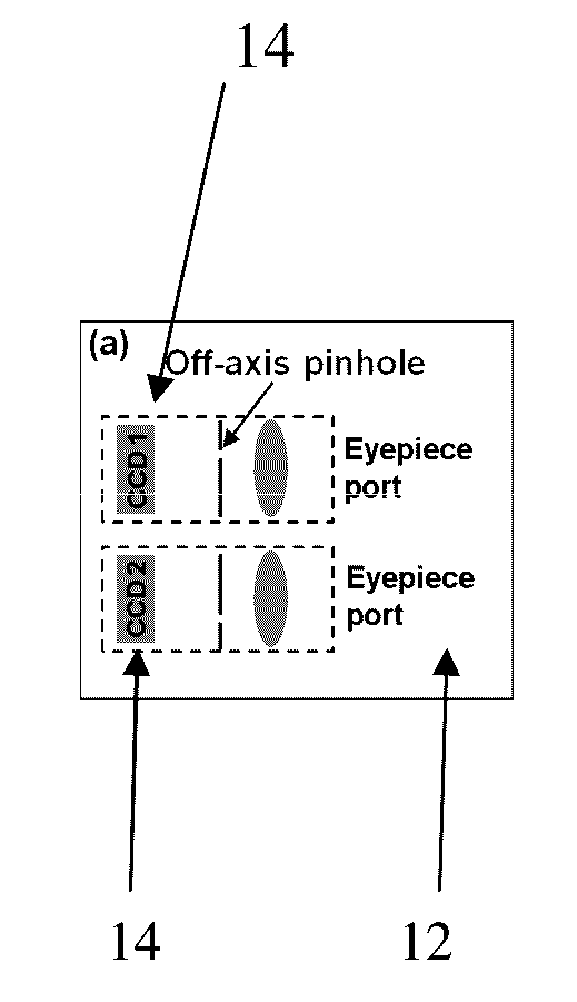
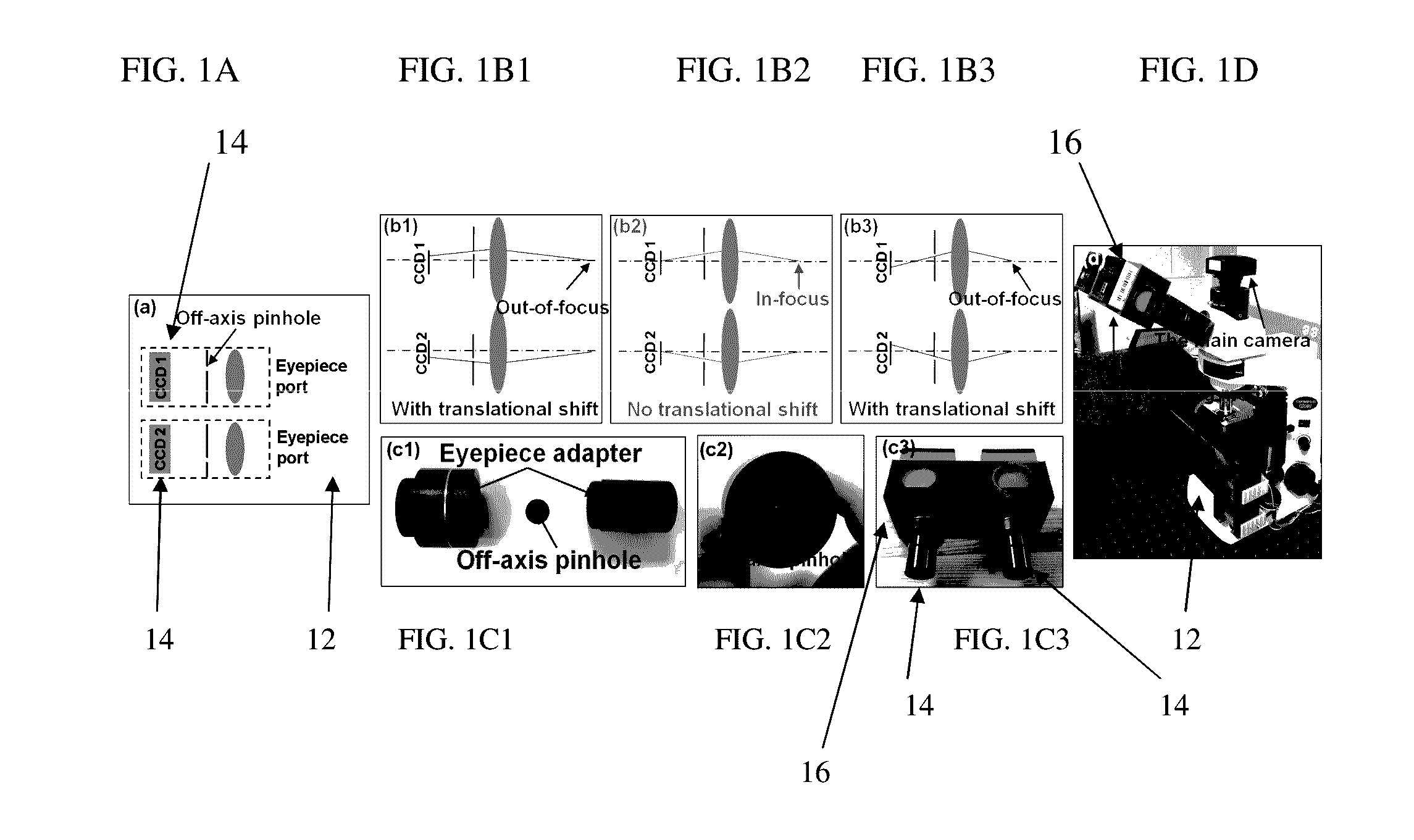
Two Cameras for Auto-Focusing and Each Camera is Modulated By a One-Pinhole Mask

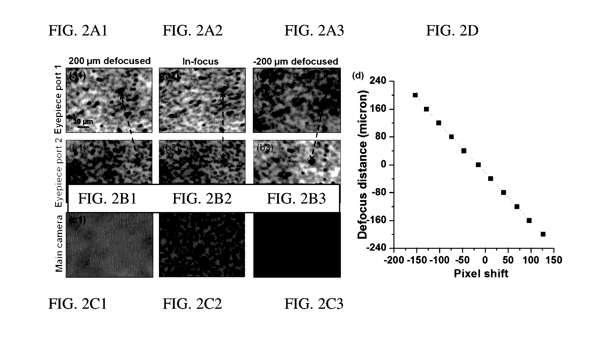
[0057]In some embodiments, one can attach two pinhole-modulated cameras 14 at the eyepiece ports of a microscope 12 for instant focal plane detection, as shown in FIG. 1A. By adjusting the positions of each pinhole of each camera 14, one can effectively change the sample view angle for the pinhole-modulated cameras 14. When the sample of the microscope 12 is placed at the in-focus position, the two captured images from cameras 14 will be identical (FIG. 1B2). When the sample is out of focus (FIGS. 1B1 and 1B3), the sample images will be projected at two different view angles, causing a translational shift in the two images. The translation shift is proportional to the defocus distance of the sample. Therefore, by identifying the translational shift of the two captured images, one can recover the optimal focal position of the sample (e.g., the in-focus position of the sample of microscope 12) without a z-...

example 2

A Camera for Auto-focusing and This Exemplary Camera is Modulated By a Two-Pinhole Mask

[0083]In another embodiment and as shown in FIG. 7, one can attach one camera 114 to a microscope. Exemplary camera 114 is attached / mounted at the epi-illumination arm of the microscope for auto-focusing.

[0084]For camera 114 and as depicted in FIG. 7, one can insert a two-pinhole mask having two pinholes at the Fourier plane of camera 114 (the auto-focusing module 114). Therefore, a captured image from this camera 114 contains two image copies of the sample, as shown in FIG. 8A (and as shown in FIG. 8B; and as shown in FIG. 8C).

[0085]If the sample is placed at the in-focus position, these two copies from one image do not have a lateral distance between them. If the sample is out-of-focus (e.g., FIG. 8A), these two image copies contained in the single image has a lateral translational shift (e.g., the distance between the two arrows in FIG. 8A). Based on this lateral translational shift, one can re...


PUM


Abstract

Advantageous instruments, assemblies and methods are provided for undertaking imaging techniques (e.g., microscopic imaging techniques). The present disclosure provides improved imaging techniques, equipment and systems. More particularly, the present disclosure provides advantageous microscopy/imaging assemblies with rapid sample auto-focusing (e.g., microscopy/imaging assemblies having instant focusing for rapid sample imaging with auto-focusing). The present disclosure provides for high-throughput whole slide imaging with instant focal plane detection. A whole slide imaging platform/assembly that uses instant focusing systems/methods for high-speed sample autofocusing is provided. Such exemplary platforms/assemblies can be used for digital pathology or the like, and can provide improved, faster and cheaper diagnosis/prognosis of ailments/diseases. At least two exemplary rapid-focus systems for whole slide imaging are provided, a first system including two pinhole-modulated cameras mounted on the eyepiece ports of a microscope platform/assembly, and a second system including one pinhole-modulated camera mounted on the epi-illumination arm for auto-focusing.

Description

CROSS-REFERENCE TO RELATED APPLICATIONS[0001]This application claims priority to Provisional Application Ser. No. 62 / 197,283 entitled “Auto-Focus System For Whole Slide Imaging” which was filed Jul. 27, 2015, the contents of which is hereby incorporated by reference in its entirety.FIELD OF THE DISCLOSURE[0002]The present disclosure relates generally to the field of imaging techniques, equipment and systems and, more particularly, to microscopy / imaging assemblies with rapid sample auto-focusing (e.g., microscopy / imaging assemblies having instant focusing for rapid sample imaging with auto-focusing).BACKGROUND OF THE DISCLOSURE[0003]In general, a whole slide imaging (“WSI”) system can be an important tool for biomedical research and clinical diagnosis. In particular, advances of computer and image sensor technologies have accelerated the development of WSI systems for high-content screening, telemedicine, and digital pathology. An important aspect of WSI systems is to maintain the sa...

Claims


Application Information

Patent Timeline
Patent Type & Authority Applications(United States)
IPC IPC(8): G02B21/00H04N5/232G02B21/20G06T7/00G02B21/36H04N5/225H04N5/247
CPCG02B21/0084H04N5/2252H04N5/23212H04N5/247H04N5/2256G06T2207/30004G06T7/0012G02B21/367G02B21/361G02B21/362G02B21/20G06T7/004H04N23/51H04N23/45H04N23/672H04N23/673H04N23/56H04N23/90
Inventor ZHENG, GUOAN
Owner UNIV OF CONNECTICUT
Why Eureka
Try Eureka