Aorticorenal Ganglion Detection

a technology of aorticorenal ganglion and ganglion, which is applied in the direction of catheters, nervous system evaluation, therapy, etc., can solve the problems of affecting affecting the function of the kidney, and reducing or cesing the activity of the kidney nerve involved in the development of hypertension, so as to prevent the shut down of the high impedance electrosurgical generator, reduce the damage to the endothelial tissue, and reduce the damage to the kidney

Inactive Publication Date: 2017-04-06
HALCYON MEDICAL INC
View PDF5 Cites 53 Cited by
  • Summary
  • Abstract
  • Description
  • Claims
  • Application Information

AI Technical Summary

Benefits of technology

[0024]Another embodiment of the present invention is directed to a catheter assembly including a multi-electrode bipolar radiofrequency electrode element located at the distal end of said catheter. Use is similar to monopolar catheter but does not require the use of dispersive electrode pad. Another embodiment of the present invention is directed to radiofrequency electrode element with a cooling feature at the distal end of said catheter. Cooling the RF electrode element during activation has several benefits including limiting endothelial tissue damage to the vessel wall and creating deeper tissue modification (e.g. deeper lesions) if desired. Cooling the RF electrode element allows for higher temperatures thus deeper lesions by preventing high impedance electrosurgical generator shut down which occurs when blood coagulation collects on the higher temperature electrode elements. Cooling mechanism may incorporate a peltier effect device, cooled fluid or gas circulated in catheter distal tip. One example of cooling the electrode element involves flushing saline through catheter body and out of through-holes manufactured into the electrode element into the blood stream during radiofrequency energy activation, thus cooling the hotter electrode element with the cooler fluid through heat transfer.

Problems solved by technology

The method includes percutaneous modification of the aorticorenal ganglion and / or postganglionic renal nerves which results in a decrease or cessation of kidney nerve activity involved in the development of hypertension.
Activation modifies (e.g. ablates when radiofrequency energy is employed) the ganglion, creating disruption of nerve signals leading to the kidney.

Method used


Image

  • Aorticorenal Ganglion Detection
  • Aorticorenal Ganglion Detection
  • Aorticorenal Ganglion Detection

Examples


experiment 1

[0127]Chronic swine study was performed to demonstrate reduction in renal nerve activity after modification of the aorticorenal ganglia. The domestic swine model is an established model for the renal system because the pig renal anatomy, including circulatory and nervous system, is similar to that of humans.

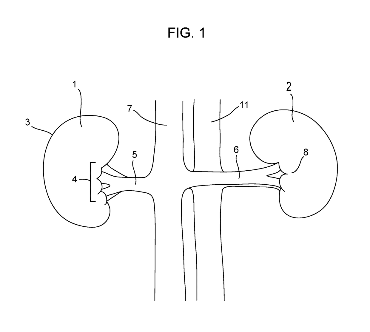
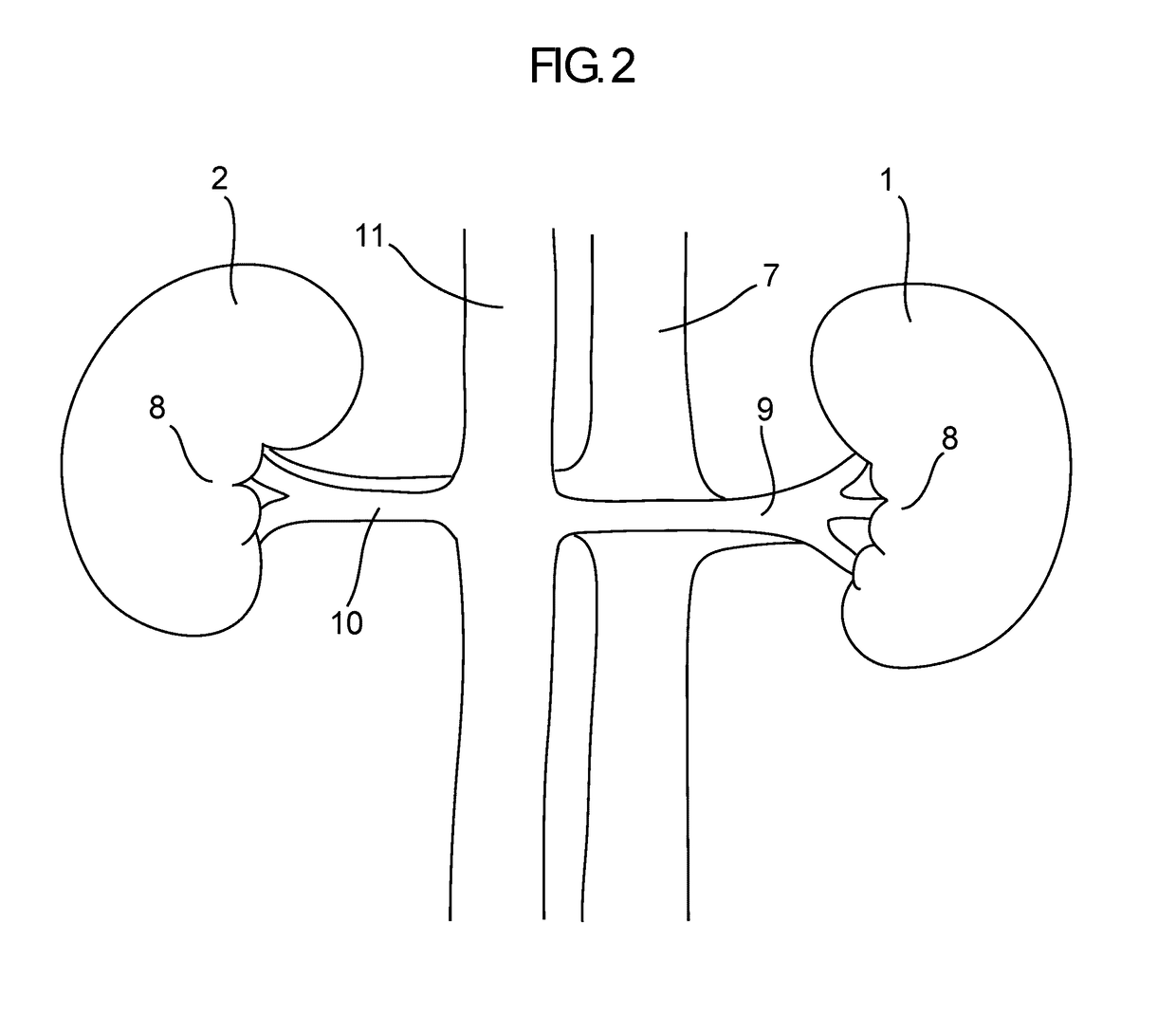
[0128]The procedure involved placing the anesthetized test animal in dorsal recumbency, followed by a 10-cm midline abdominal incision in order to access the renal anatomy. Peritoneum was removed to expose left and right renal artery, vein, aorta and vena cava. Adventitia was stripped from the renal arteries and veins to expose the renal nerve plexus and aorticorenal ganglia. Direct electrical stimulation of the ganglia was performed at 15 volts, 5 Hz and 0.5 msec. pulse duration using a Grass Instruments SD9 Square Pulse Stimulator (Grass Technologies, Warwick, R.I.). Proper identification of the aorticorenal ganglion was confirmed during stimulation by observation of renal arte...

experiment 2

[0130]An acute swine study was performed to evaluate the feasibility of detecting an acute physiological response to percutaneous stimulation of the aorticorenal ganglion and renal nerve tissue. The procedure involved creating percutaneous access to the renal venous and arterial vasculature through a jugular and femoral puncture site of an anesthetized test animal in dorsal recumbency. Guide sheaths used for radiopaque contrast delivery were placed with fluoroscopic guidance in both the left renal vein and left renal artery to perform a baseline nephrogram. FIG. 17 is a frame capture of the baseline nephrogram showing normal intrarenal vessel emptying and kidney perfusion.

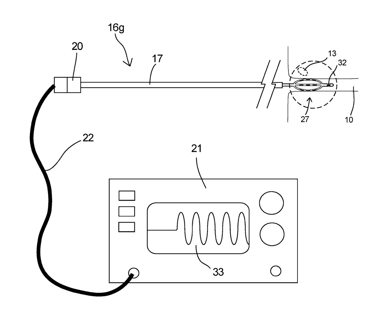
[0131]A modified electrophysiology catheter (5 French Marinr™ Ablation Catheter, Medtronic, Minneapolis, Minn.) attached to a Grass Instruments SD9 Square Pulse Stimulator (Grass Technologies, Warwick, R.I.) was percutaneously placed in the left renal vein. Direct stimulation of the renal vein wall was performed at...

experiment 3

Section

[0314]A chronic swine study was performed to demonstrate a reduction in renal nerve activity after percutaneous catheter modification of the aorticorenal ganglia (ARG).

[0315]Experiment 3 was similar to Experiment 1 with the exception that the ARG was modified intravascularly by percutaneous catheter RF ablation at a single site (i.e. Target Location) within the renal artery.

[0316]The study involved three treated and two naïve swine. Urine and blood panel analysis were performed at each stage of the study, pre- / post-treatment and pre-kidney tissue harvest, and nephrograms were performed for the treated swine pre-treatment and pre-kidney tissue harvest. A veterinarian managed assessment of animal health throughout the study based on this data and assured proper pharmacology and diet were applied.

[0317]The procedure involved creating percutaneous access to the renal arterial vasculature through a femoral puncture site of an anesthetized test animal in dorsal recumbency. Guide ca...


PUM


Abstract

Devices and methods that regulate the innervation of the kidney by detection and modification of the aorticorenal ganglion. Devices for percutaneous detection and treatment of the aorticorenal ganglion via a blood vessel to modify renal sympathetic activity.

Description

RELATED APPLICATIONS[0001]This application claims priority to U.S. Provisional Application Ser. No. 62 / 237,966 filed Oct. 6, 2015 entitled Aorticorenal Ganglion Detection, which is hereby incorporated herein by reference in its entirety.BACKGROUND OF THE INVENTION[0002]Hypertension or abnormally high blood pressure is a growing public health concern for which successful treatment often remains elusive. Sixty-seven million Americans—about one-third of the adult population—have high blood pressure and these numbers are increasing as the population ages and obesity accelerates.[0003]Hypertension is more common in men than women and afflicts approximately 50% of the population over the age of 65. Hypertension is serious because people with the condition have a higher risk for heart disease and other medical problems than people with normal blood pressure. If left untreated, hypertension can lead to arteriosclerosis, heart attack, stroke, enlarged heart and kidney damage.[0004]Blood pres...

Claims


Application Information

Patent Timeline
Patent Type & Authority Applications(United States)
IPC IPC(8): A61B18/14A61B18/18A61N7/02A61B18/12A61B18/24
CPCA61B18/1492A61N2007/003A61B18/24A61N7/02A61B18/1815A61B2018/1253A61B2018/126A61B2018/00511A61B2018/00404A61B2018/00434A61B2018/0022A61B2018/00267A61B2018/143A61B2018/00577A61B2018/00589A61B18/1206A61B5/40A61B5/6852A61B5/6886A61B2018/0016A61B2018/00214A61B2018/00702A61B2018/00791A61B2018/00839A61B2018/00875A61B2018/1425A61B2018/1435A61N1/0551A61N1/0558A61N1/36007A61N1/36017
Inventor HARRINGTON, DOUGLAS CHRISTOPHERTHOMAS, MARKHOLT, WILLIAM DAVID
Owner HALCYON MEDICAL INC
Why Eureka
Try Eureka