Medical image analysis for identifying biomarker-positive tumor cells

a tumor cell and image analysis technology, applied in image analysis, image enhancement, instruments, etc., can solve the problems that human observers cannot perform quantitative analysis of whole slides, such as counting target objects, or quantitation of slides, and achieve the effect of stable segmentation results

Active Publication Date: 2017-04-13
VENTANA MEDICAL SYST INC
View PDF3 Cites 79 Cited by
  • Summary
  • Abstract
  • Description
  • Claims
  • Application Information

AI Technical Summary

Benefits of technology

[0014]Said features may be advantageous as a highly accurate method of identifying biomarker-positive tumor cells may be provided. Instead of identifying the tumor cells in a single image analysis step, the nuclei and the cell membranes are identified and used as a basis for identifying complete cells. This may increase accuracy because more characteristic features of nuclei and membranes may be evaluated and image analysis algorithms may be used which are specially adapted to identifying nuclei or cell membranes. In addition, instead of identifying the nuclei and the cell membranes independently of each other, the process of identifying the cell membranes also takes into consideration positional information of the already identified nuclei. This may significantly increase accuracy, because staining artifacts whose shape or other property is similar to a cell membrane can be identified as artifacts if, for example, said staining artifacts do not lie within a maximum distance from one of the identified nuclei. Using positional information of the identified nuclei may thus increase the accuracy of cell membrane identification, which again may increase accuracy of biomarker-positive tumor cell identification.
[0089]Calculating a membrane completeness score may help to evaluate and estimate the accuracy of the tumor cell identification. The watershed approach, for example, may automatically create and extend “artificial dams” along identified watershed lines which may cover even regions of the slide where no biomarker signal could be observed. By calculating the completeness score for such a tumor cell, it may be determined that actually the signal is too weak to safely consider said tumor cell as a biomarker-positive cell. Such tumor cells may be identified, by the completeness score calculation step, as a tumor cell with an incomplete or absent cell membrane. Such a tumor cell may be re-categorized as a biomarker-negative tumor cell. In addition, a tumor cell having an incomplete or absent cell membrane may be identified as a hint that the tumor cells do not express the biomarker or express the biomarker in a very limited amount. In addition, calculating a completeness score may allow assessing the quality of any calculated intensity based score.

Problems solved by technology

Quantitative analysis of a whole-slide, such as counting target objects such as cells of a certain kind, or the quantitation of a staining response for all cells on a slide, is not feasible for human observers.
Abnormal MET activation in cancer correlates with poor prognosis, where aberrantly active MET triggers tumor growth, angiogenesis and metastasis.
Manual assessment of these criteria is difficult or impossible, similar to detection and scoring of membranous and cytoplasmic regions in other IHC (“immunohistochemistry”)-stained tissue slides, for assays such as HER2 and EGFR, and for other cancerous tissue types, such as breast and gastric cancers.
Oversegmentation can be serious enough to render the results of the algorithm virtually useless.

Method used


Image

  • Medical image analysis for identifying biomarker-positive tumor cells
  • Medical image analysis for identifying biomarker-positive tumor cells
  • Medical image analysis for identifying biomarker-positive tumor cells

Examples


Embodiment Construction

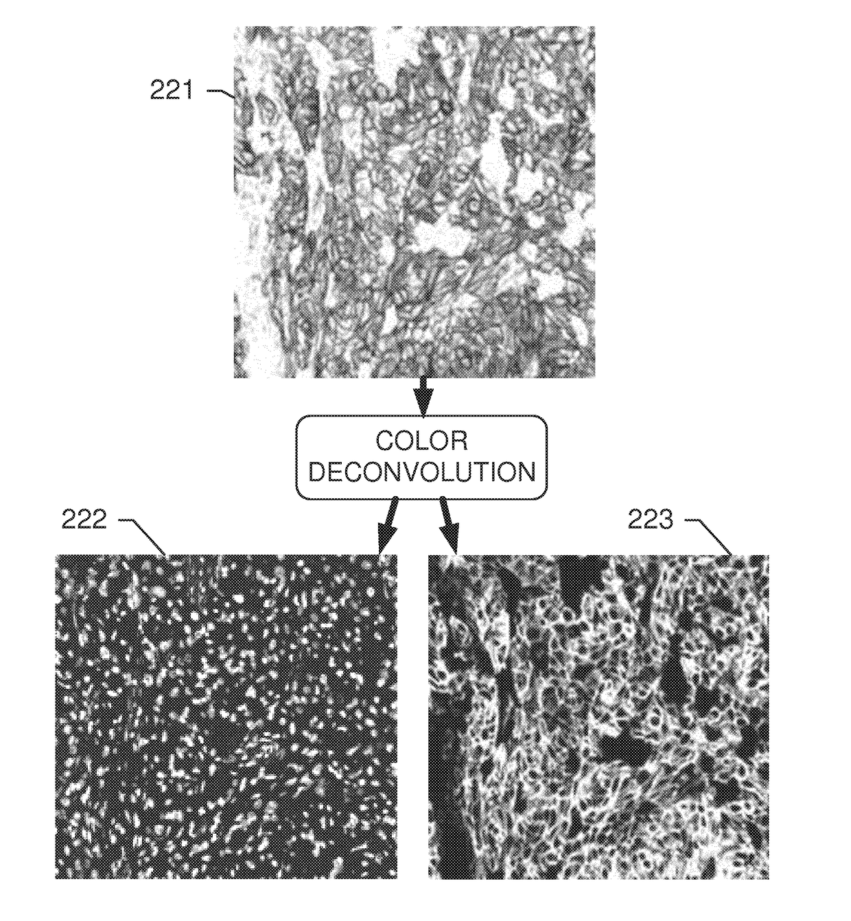
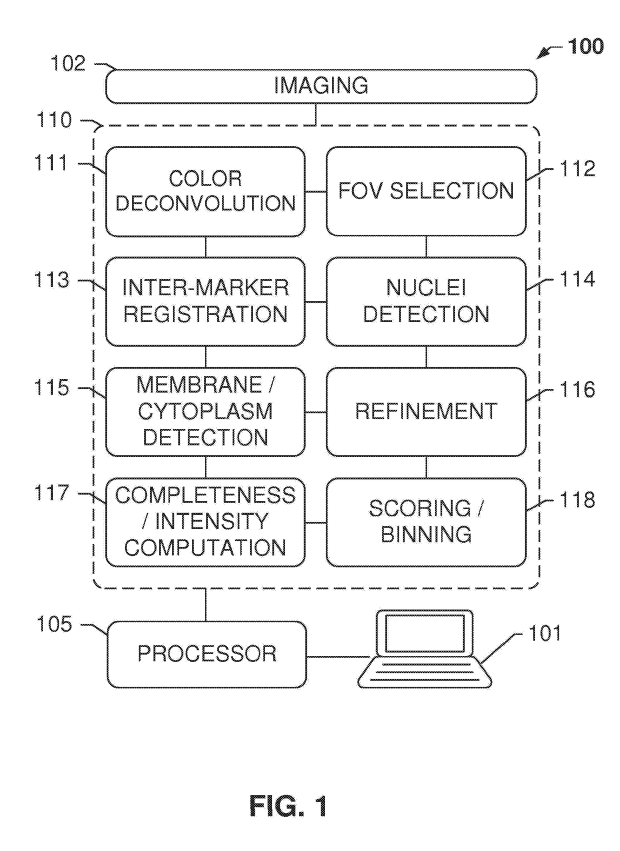
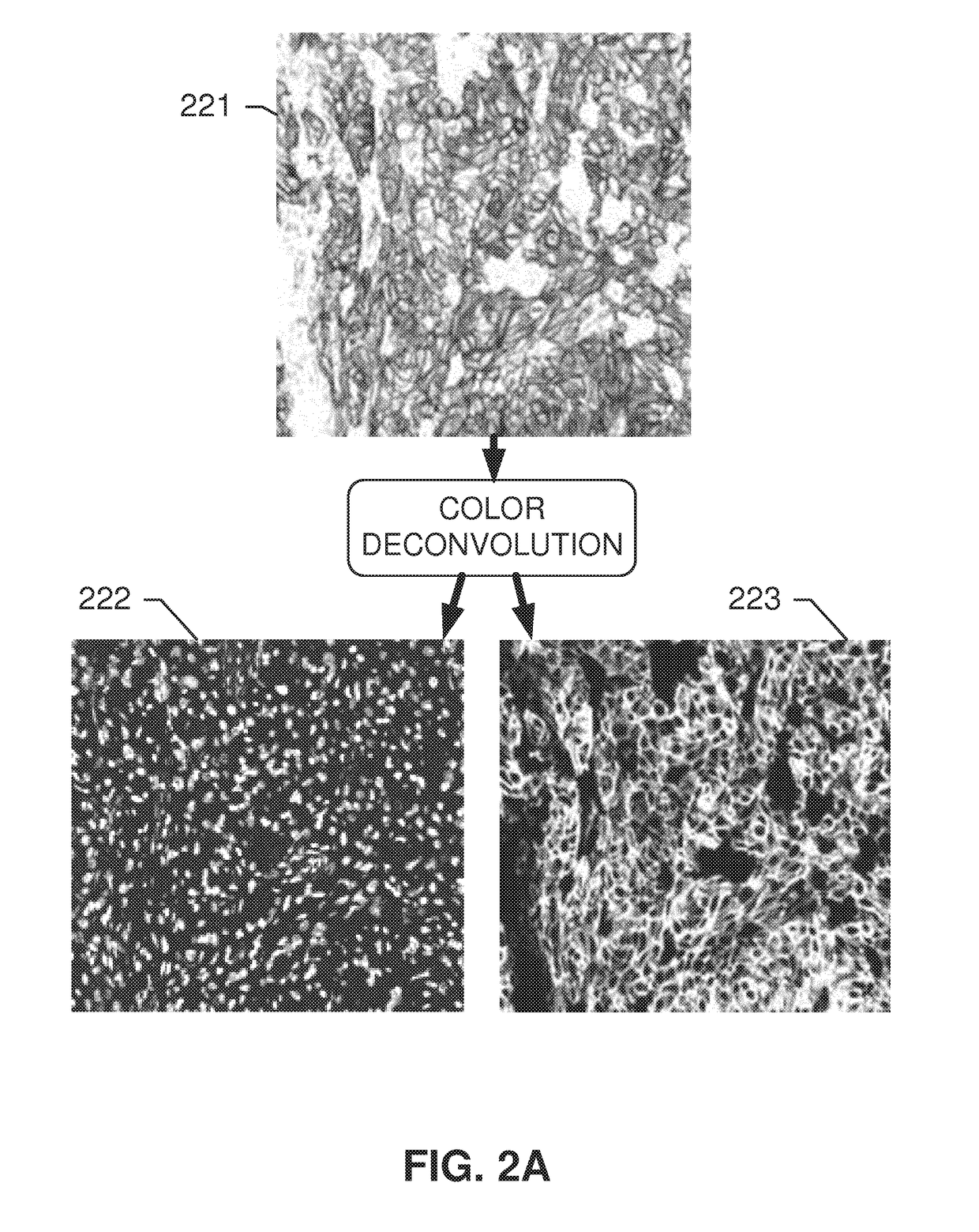
[0148]Systems and methods disclosed herein relate to an automated image analysis algorithm and workflow to score digitized slides, having biological specimen or specimens thereon, stained with IHC assays which stain the membranous and cytoplasmic regions. The present invention is described, for exemplary purposes, in connection whole NSCLC slides that are stained with c-MET IHC assay. However, the disclosed operations may be applicable to any other combinations of membranous and cytoplasmic stains and nuclei counterstains as will be evident to persons having ordinary skill in the art in light of this disclosure.

[0149]A “counterstain” can be, for example, a stain for staining nuclei. For example, the counterstain can be of a contrasting color in respect to the stain used to color the membrane and cytosolic components comprising the biomarker. According to embodiments, the nuclei are made visible only by the counterstain, not by the stain used for staining the biomarker. According to ...


PUM


Abstract

A method for identifying biomarker-positive tumor cells is disclosed. The method includes, for example, reading a first digital image and a second digital image into memory, the first and second digital image depicting the same area of a first slide; identifying a plurality of nuclei and positional information of said nuclei by analyzing the light intensities in the first digital image; identifying cell membranes which comprise the biomarker by analyzing the light intensities in the second digital image and by analyzing the positional information of the identified nuclei; and identifying biomarker-positive tumor cells in said area, wherein a biomarker-positive tumor cell is a combination of one identified nucleus and one identified cell membrane that surrounds the identified nucleus.

Description

CROSS-REFERENCE TO RELATED APPLICATIONS[0001]This patent application is a continuation of International Patent Application No. PCT / EP2015 / 053757 filed Feb. 23, 2015, which claims priority to and the benefit of U.S. Provisional Application No. 61 / 943,271, filed Feb. 21, 2014. Each patent application is incorporated herein by reference as if set forth in its entirety.FIELD OF THE INVENTION[0002]The present subject disclosure relates to imaging for medical diagnosis. More particularly, the present subject disclosure relates to automatically identifying biomarker-positive tumor cells on a slide.BACKGROUND AND RELATED ART[0003]In the field of digital pathology, biological specimens such as tissue sections, blood, cell cultures and the like may be stained with one or more stains and analyzed by viewing or imaging the stained specimen. Observing the stained specimen, in combination with additional clinical information, enables a variety of processes, including diagnosis of disease, prognos...

Claims


Application Information

Patent Timeline
Patent Type & Authority Applications(United States)
IPC IPC(8): G06T7/00
CPCG06T7/0012G06T2207/10004G06T2207/30024G06T2207/30096G06T2207/30242G06V20/695
Inventor CHUKKA, SRINIVASYUAN, QUAN
Owner VENTANA MEDICAL SYST INC
Why Eureka
Try Eureka