
Premature neonate life support environmental chamber for use in mri/nmr devices

Active Publication Date: 2017-07-06
ASPECT IMAGING
View PDF8 Cites 4 Cited by
  • Summary
  • Abstract
  • Description
  • Claims
  • Application Information

AI Technical Summary

Benefits of technology

The present invention provides an MRI-safe cart with a telescopic or folding mechanism that allows for different lengths of the cart column. This feature makes it easier to move and position the cart in different spaces while still maintaining safety standards. Additionally, the reversible collapsible column can also be expanded and contracted with the aid of a telescopic or folding mechanism, allowing for even more flexibility in usage.

Problems solved by technology

A well-known technical problem in the design of an MRI / NMR device is that of the fringing magnetic fields.
These fringing magnetic fields can cause interference with nearby devices and lead to safety concerns arising from the possibility of placing magnetic material in propinquity to the MRI / NMR device as well as limiting the proximity of two MRI / NMR devices or other devices.
This limitation results in the fact that almost invariably the MRI / NMR device is stored in a special isolated facility.
This last problem is of particular concern for cases in which it is required to take MRI imaging examinations and measurements of a premature neonate without transferring the premature neonate between a premature neonate intensive-care ward and an MRI imaging facility, without decoupling and disconnecting the premature neonate from life-support systems.
The above-cited references do not provide a system or method enabling a premature neonate to undergo an MRI / NMR examination e.g., in the premature neonate intensive-care ward and require that the premature neonate be detached from the life-supporting systems during transfer to the MRI-compatible incubator.
Thus, during placing the patient, such as a neonate, neonate in the prior art incubator, the patient or neonate is detached from the life-support systems, which can endanger the health of the patient.
Therefore, there is a long felt and hitherto unmet need for providing improved incubator devices.
Many accidents were reported when metallic items were pulled in by the force of the magnetic field and harmed a patient during imaging.
Other risks may be peripheral nerve stimulation, exposure to a loud noise (up to 120 dB), generated by the rapid switching of the magnetic field gradients, or overheating may occur due to absorption of the energy that is utilized to generate the magnetic spin.
Another risk involves an unintentional shut-down of a superconducting electromagnet (“quench”), resulting in the rapid boiling of liquid helium from the device.
The rapidly expanding helium if released into the scanner room may cause displacement of the oxygen and present a risk of asphyxiation.
Any transfer or movement of the baby may require the transfer or reconnection of attached medical devices, posing an additional stress on the baby.
Further, any changes in location of the neonate may expose him to infection from an unprotected environment.
The above-cited references does not provide a system enabling a premature neonate to undergo an MRI / NMR examination e.g., in the premature neonate intensive-care ward and require that the premature neonate be detached from the life-supporting systems during transfer to the MRI-compatible incubator.
Thus, during placing the patient, such as a neonate, neonate in the prior art incubator, the patient or neonate is detached from the life-support systems, which can endanger the health of the patient.
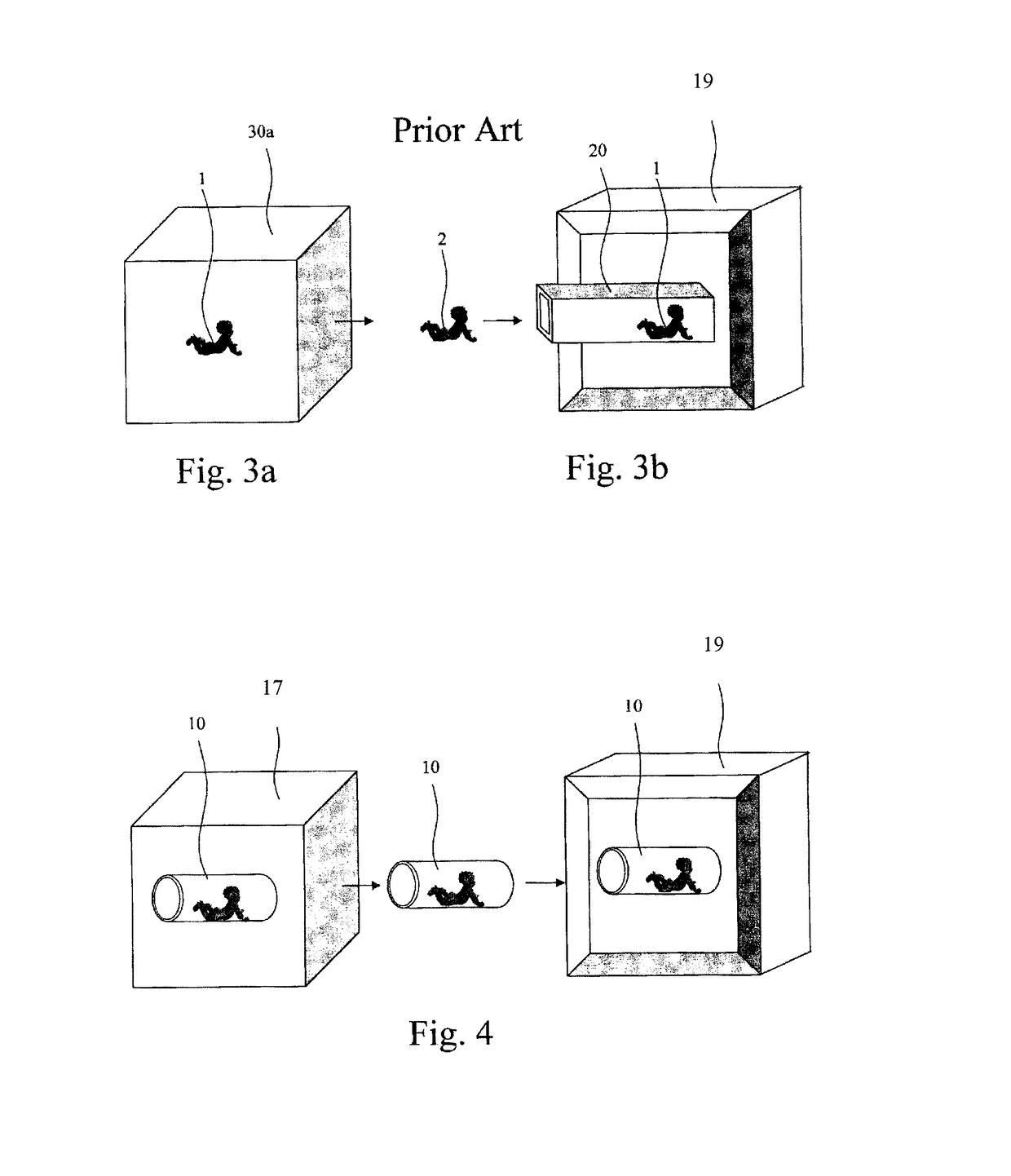
Further the above sited reference does not provide a system of an incubator as a portion of a cart accepted in an MRD bore thereby providing a single step process for imaging a neonate.

Method used


Image

  • Premature neonate life support environmental chamber for use in mri/nmr devices
  • Premature neonate life support environmental chamber for use in mri/nmr devices
  • Premature neonate life support environmental chamber for use in mri/nmr devices

Examples


Embodiment Construction

[0235]The following description is provided, alongside all chapters of the present invention, so as to enable any person skilled in the art to make use of the invention and sets forth the best modes contemplated by the inventor of carrying out this invention. Various modifications, however, will remain apparent to those skilled in the art, since the generic principles of the present invention have been defined specifically to provide a premature neonate life support environmental chamber and methods using the same.

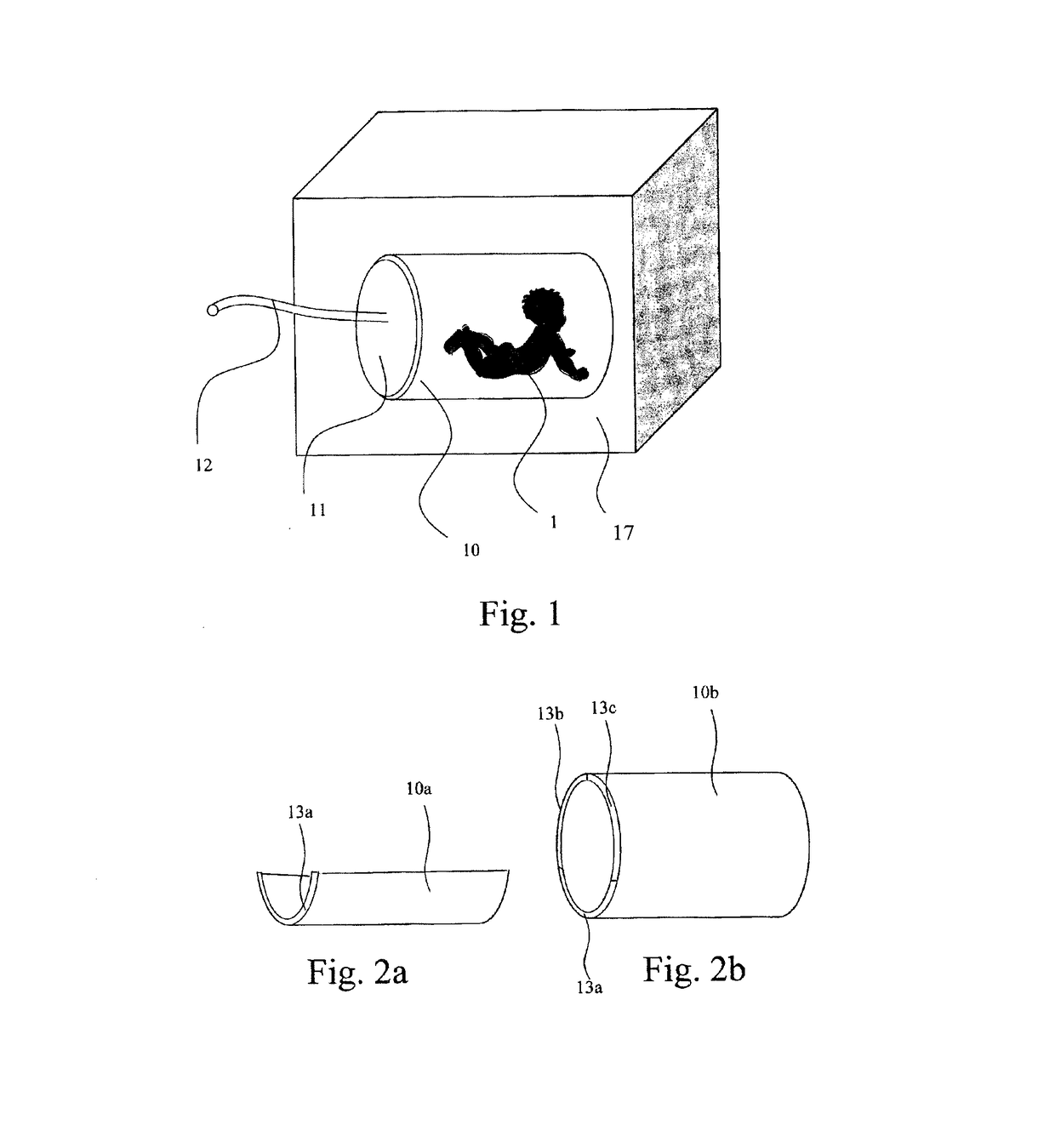
[0236]The present invention provides a premature neonate life Support Environmental Chamber (hereinafter ‘SEC’), permeable to magnetic fields for use in a portable MRI / NMR device, such as an MRD device. This environmental chamber SEC is adapted to accommodate any neonate incubator permeable to magnetic fields. Hence, the SEC is configured such that the SEC and the incubator include a closed life support system for the neonate.

[0237]The essence of the present invention is t...


PUM


Abstract

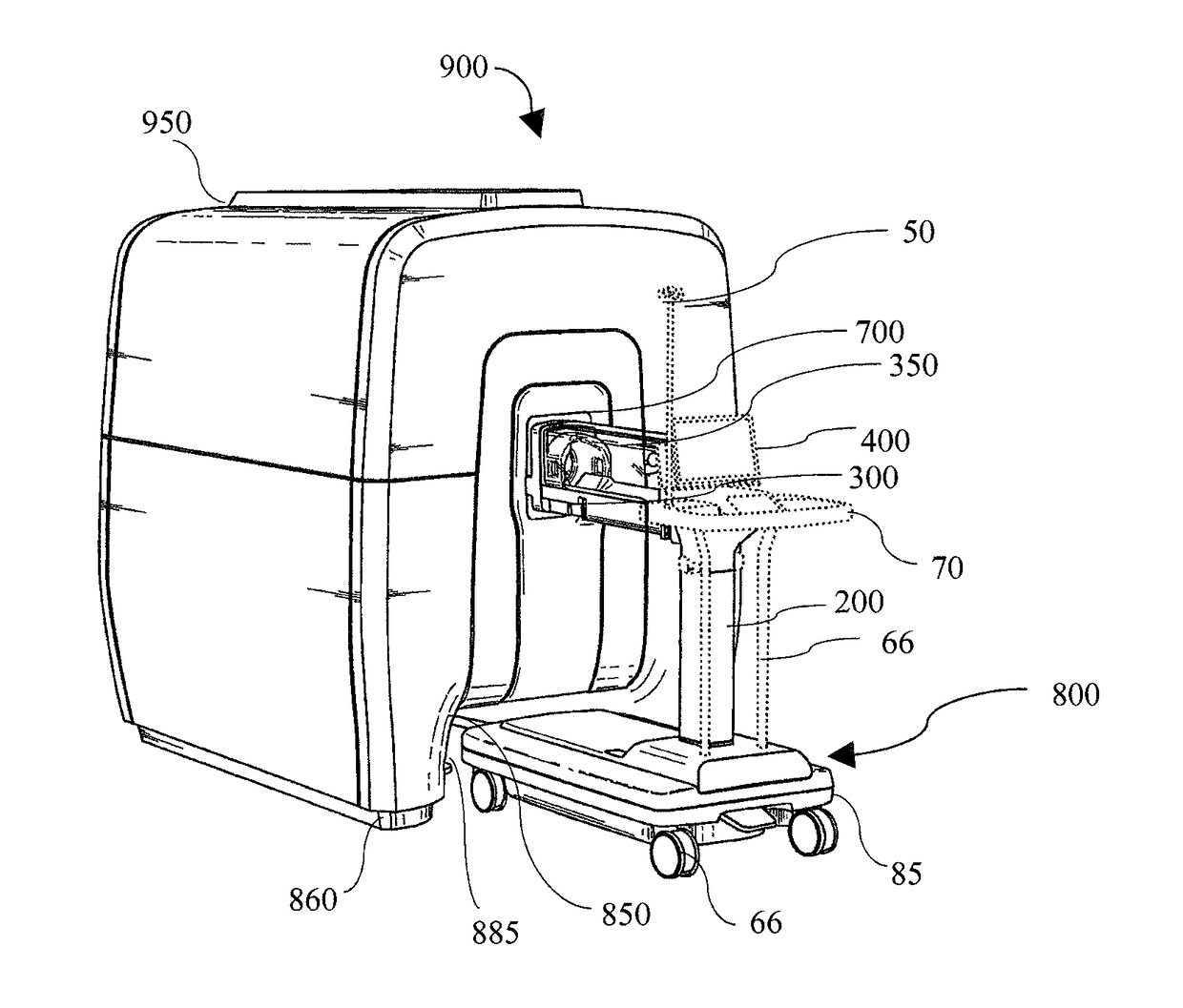
A magnetic resonance system, useful for imaging a patient, comprising: (a) a magnetic resonance device (MRD) for imaging a patient, comprising an open bore, the MRD at least partially contained in an envelope comprising in its circumference at least one recess; and, (b) an MRI-safe cart made of MRI-safe material, comprising a substantially horizontal base and at least one substantially horizontal incubator above the base, the base and the incubator are interconnected by at least one pillar. At least a portion of the cart and the MRD are configured to fit together such that at least a portion of the incubator is reversibly housed within the MRD, and further at least a portion of the base is reversibly housed within at least one recess.

Description

CROSS REFERENCE TO RELATED APPLICATIONS[0001]This application is a continuation-in-part of U.S. patent application Ser. No. 13 / 808,476, filed on Jul. 11, 2013, which is a national stage filing of PCT / IL11 / 00537, filed on Jul. 7, 2011, which claims benefit of and priority to U.S. provisional application No. 61 / 361,936, filed on Jul. 7, 2010, all of which are incorporated herein by reference in their entirety. This application is also a continuation-in-part of U.S. patent application Ser. No. 14 / 892,207, filed on Nov. 19, 2015, which is a national stage filing of PCT / IL2014 / 050450, filed on May 21, 2014, which claims the benefit of and priority to U.S. provisional patent application No. 61 / 994,901, filed on May 18, 2014, all of which are incorporated herein by reference in their entirety.FIELD OF THE INVENTION[0002]The present invention relates to equipment for MRI imaging of premature neonates. In particular, it pertains to means and methods for enabling MRI examinations and measurem...

Claims


Application Information

Patent Timeline
IPC IPC(8): A61B5/055G01R33/30A61G11/00G01R33/422
CPCG01R33/422G01R33/307A61B5/0555A61B2503/045A61G11/002A61B5/6887A61G11/00G01R33/3806A61B5/055
Inventor RAPOPORT, URI
Owner ASPECT IMAGING
Learn More