
Ultrasound imaging apparatus

a technology of ultrasound imaging and ultrasound, applied in the field of ultrasound imaging apparatus, can solve the problems of time-consuming, complicated examination, and patient risk, and achieve the effect of improving ultrasound imaging analysis and low technical effor

Inactive Publication Date: 2017-09-07
KONINKLJIJKE PHILIPS NV
View PDF3 Cites 34 Cited by
  • Summary
  • Abstract
  • Description
  • Claims
  • Application Information

AI Technical Summary

Benefits of technology

The present invention is about a method to get ultrasound data of a patient by using an ultrasound acquisition unit and transform that data to create a virtual field of view inside the patient's body. This virtual field of view can be determined based on the position of the ultrasound probe or by using anatomical context and segmenting data. This approach allows for an internal view of the patient's body without needing to insert a catheter. The technical effect is that this method reduces the effort required to provide ultrasound images from an internal view of the patient's body.

Problems solved by technology

The ultrasound catheters including ultrasound echo probes are expensive and have to be introduced into the patient's body so that the examination is complicated, time consuming and probably risky for the patient.

Method used


Image

  • Ultrasound imaging apparatus
  • Ultrasound imaging apparatus
  • Ultrasound imaging apparatus

Examples


Embodiment Construction

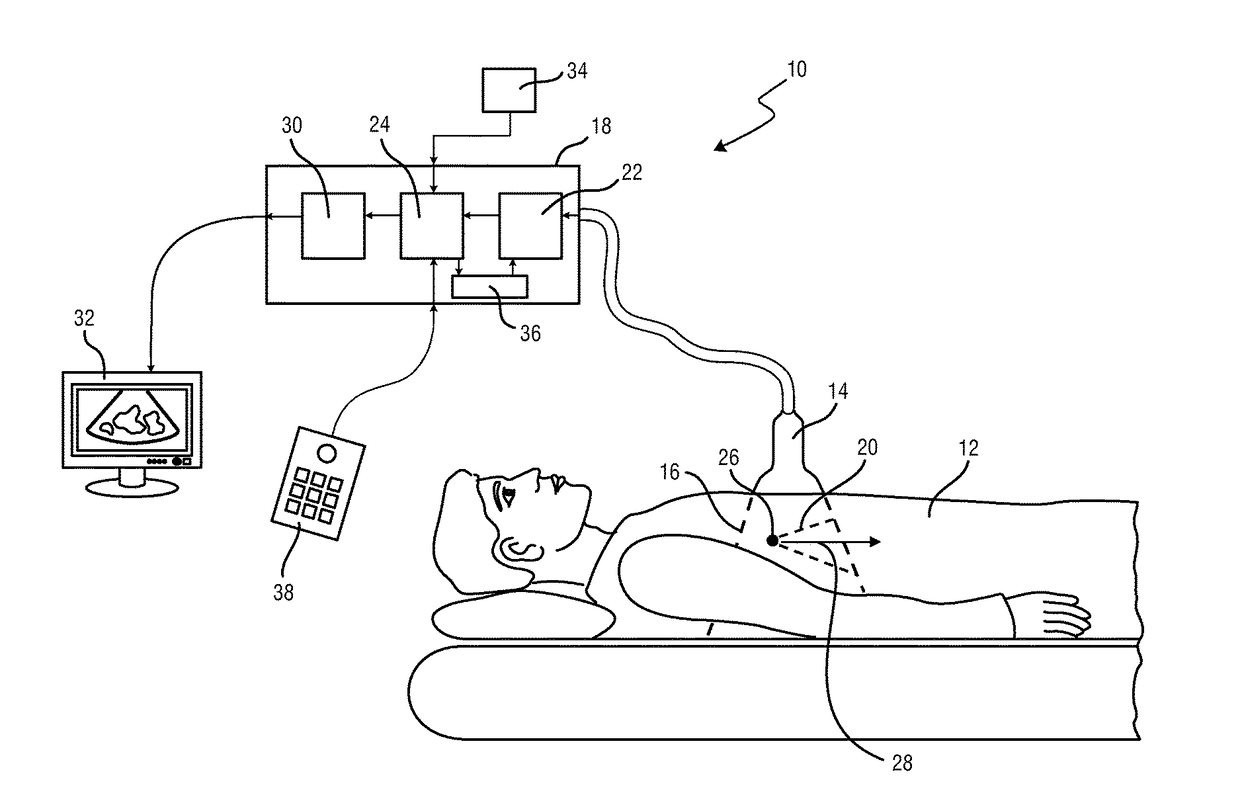
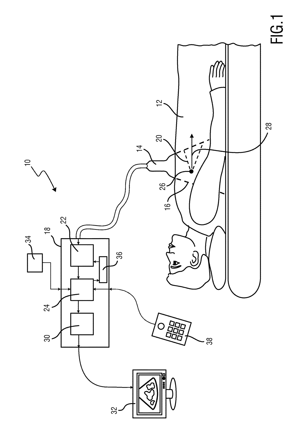
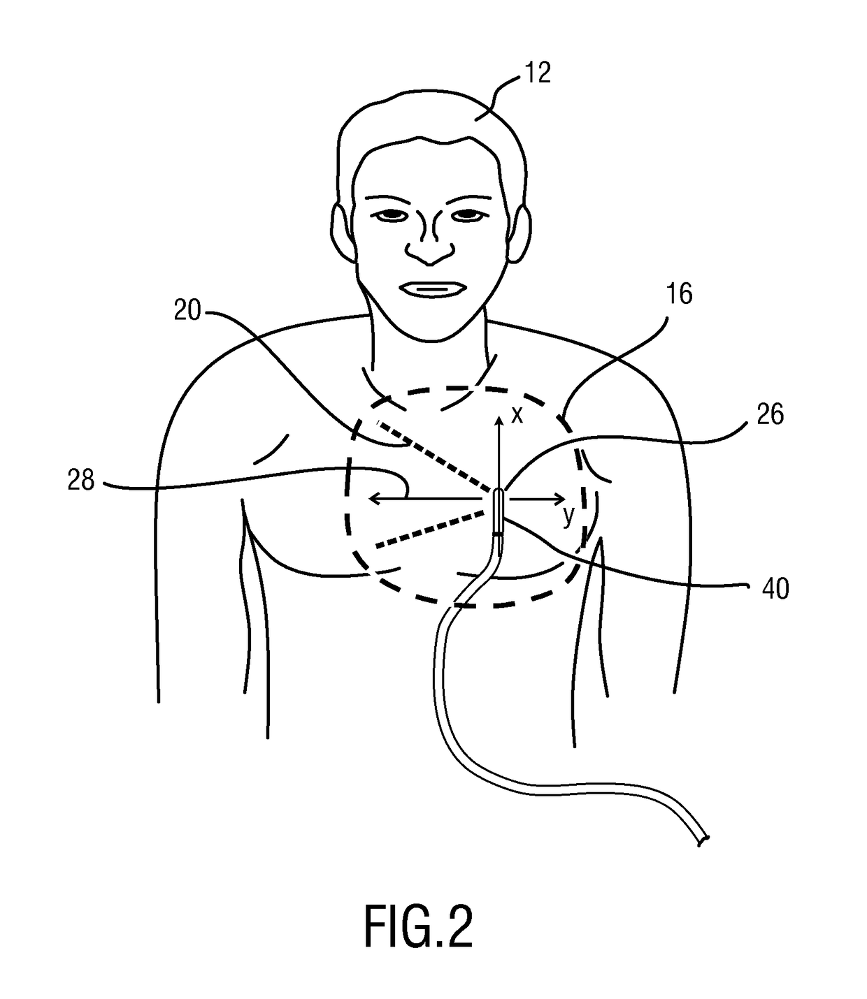
[0037]FIG. 1 shows a schematic illustration of an ultrasound imaging apparatus 10 according to one embodiment. The ultrasound imaging apparatus 10 is applied to inspect a volume of an anatomical side, in particular an anatomical side of a patient 12. The ultrasound imaging apparatus comprises an ultrasound acquisition unit 14 in particular an ultrasound probe 14 having at least one transducer array including a multitude of transducer elements for transmitting and receiving ultrasound waves. The transducer elements are preferably arranged in a 2D array for providing 3D ultrasound image data. The ultrasound acquisition unit 14 acquires ultrasound data in a field of view 16 within the patient's body and provides corresponding 3D ultrasound data.

[0038]The ultrasound imaging apparatus 10 comprises in general an image processing apparatus 18 for evaluating the ultrasound data received from the ultrasound acquisition unit 14 and for transforming the ultrasound data in the field of view 16 ...


PUM


Abstract

An ultrasound imaging apparatus (10) for providing ultrasound images of a patient (12) is disclosed. The imaging apparatus (10) comprises an ultrasound acquisition unit (14) for acquiring ultrasound data (42) of a patient's body in a field of view (16), a position determining unit (24) for determining a position (26) within the patient's body. An ultrasound data transformation unit (30) is provided for transforming the ultrasound data in the filed of view on the basis of the determined position to transformed ultrasound data (42) in a virtual field of view (20) having a virtual viewing direction (28) different from the viewing direction of the ultrasound acquisition unit.

Description

FIELD OF THE INVENTION[0001]The present invention relates to an ultrasound imaging apparatus for providing ultrasound images of a patient. The present invention further relates to an ultrasound imaging method for providing ultrasound images of a patient and a computer program comprising program code means for causing a computer to carry out steps of the method when said computer program is carried out on a computer.BACKGROUND OF THE INVENTION[0002]In the field of medical imaging systems it is generally known to use catheters including ultrasound echo probes for providing an ultrasound view from a position within the patient's body, such as e.g. intracardiac echocardiography. A corresponding ultrasound catheter echo probe for providing intracardiac ultrasound images is e.g. known from U.S. Pat. No. 8,270,694 B2.[0003]The ultrasound catheters including ultrasound echo probes are expensive and have to be introduced into the patient's body so that the examination is complicated, time co...

Claims


Application Information

Patent Timeline
IPC IPC(8): A61B6/12G06T7/00A61B8/00A61B6/00A61B8/12A61B8/08
CPCA61B6/12A61B8/12A61B8/08A61B8/4245G06T7/0012A61B8/0883A61B6/466A61B8/523A61B8/483A61B8/085A61B8/467
Inventor WEBER, FRANK MICHAELSTEHLE, THOMAS HEIKOWAECHTER-STEHLE, IRINAWEESE, JUERGEN
Owner KONINKLJIJKE PHILIPS NV
Learn More