
Medical image processing apparatus and medical image processing method

a medical image processing and image processing technology, applied in image enhancement, angiography, instruments, etc., can solve the problems of failure of the heart, large calculation time, and time also needed

Active Publication Date: 2018-01-18
CANON MEDICAL SYST COPRPORATION
View PDF11 Cites 1 Cited by
  • Summary
  • Abstract
  • Description
  • Claims
  • Application Information

AI Technical Summary

Benefits of technology

The patent text describes a medical image processing apparatus or method for assessing the degree of progress of stenosis (a narrowing of a blood vessel) in the heart caused by ischemic heart disease. The invention aims to provide a non-invasive and efficient way to measure the degree of progress of stenosis and risk assessment, which is currently done through an invasive method using a pressure wire. The invention proposes a simulation-based FFR (Fractional Flow Reserve) estimation method using fluid analysis, which reduces the time required for the simulation and allows for quicker and more accurate assessment of the degree of progress of stenosis and risk assessment.

Problems solved by technology

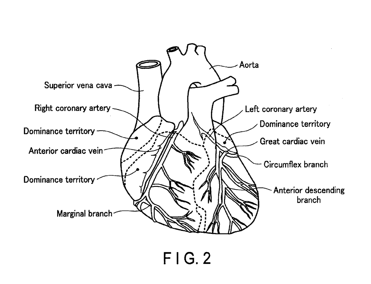
In general, in ischemic heart diseases, a blood flow to the cardiac muscle is hindered due to occlusion, stenosis, etc. of a coronary artery, and the supply of blood becomes deficient or stops, leading to a failure of the heart.
However, since the measurement of the pressures Pin and Pout with use of the pressure wire is invasive, there is a demand for a non-invasive measuring method and FFR estimation method.
A problem with such 3D simulation is that a great deal of calculation time is required.
Thus, such a problem arises that time is also needed until selecting a therapeutical method by using the FFR, and the 3D simulation is not suitable when there is no time to lose.
At present, however, it is not possible to assess the causal relationship between an ischemic cardiac muscle and a stenosed part to be treated, or to make risk assessment.
Specifically, the FFR fails to be properly reflected on the assessment of the causal relationship between an ischemic cardiac muscle and a stenosed part to be treated, and on the risk assessment.
Thus, such a problem arises that there is a concern of occurrence of a human error, such as unnecessary treatment or an oversight.

Method used


Image

  • Medical image processing apparatus and medical image processing method
  • Medical image processing apparatus and medical image processing method
  • Medical image processing apparatus and medical image processing method

Examples


first embodiment

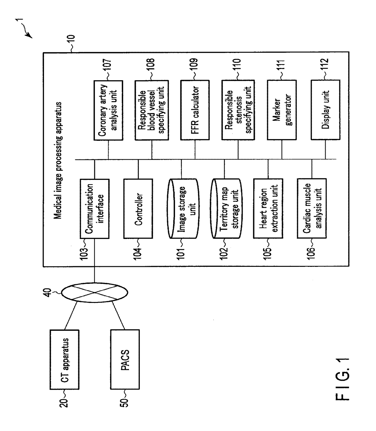
[0040]A medical image processing apparatus disclosed by this embodiment comprises a first extraction unit, a calculation unit, a second extraction unit, a specifying unit and a display. The first extraction unit is implemented by processing circuitry and configured to extract a plurality of coronary arteries depicted in data of images of a plurality of time phases relating to the heart, and to extract at least one stenosed part depicted in each of the extracted coronary arteries. The calculation unit is implemented by processing circuitry and configured to calculate a pressure gradient of each of the extracted coronary arteries, based on tissue blood flow volumes of the plurality of extracted coronary arteries. The second extraction unit is implemented by processing circuitry and configured to extract an ischemic region depicted in the images. The specifying unit is implemented by processing circuitry and configured to specify a responsible blood vessel of the ischemic region by ref...

second embodiment

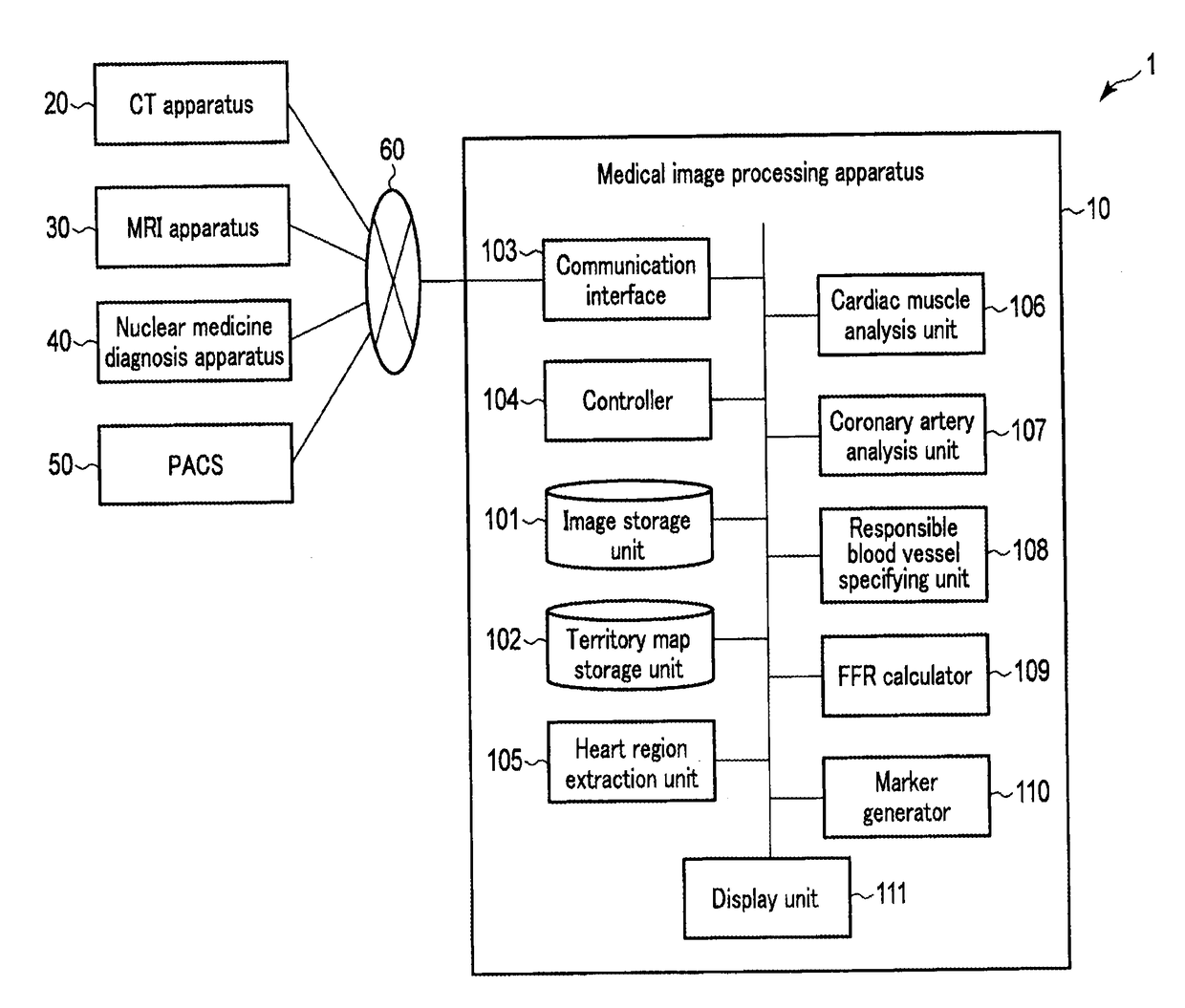
[0071]FIG. 11 is a schematic view illustrating a configuration example of a medical image processing system including a medical image processing apparatus according to a second embodiment. A medical image processing system 1 illustrated in FIG. 11 is a system in which a medical image processing apparatus 10, a CT (Computed Tomography) apparatus 20, an MRI (Magnetic Resonance Imaging) apparatus 30, a nuclear medicine diagnosis apparatus 40, and a PACS (Picture Archiving and Communication System) 50 are communicably connected via a network 60 such as a LAN (Local Area Network) or a public electronic communication network. Thus, the medical image processing apparatus 10 is provided with a communication interface 103 which enables communication with the CT apparatus 20, MRI apparatus 30, nuclear medicine diagnosis apparatus 40 and PACS 50.

[0072]As illustrated in FIG. 11, the medical image processing apparatus 10 includes an image storage unit 101, a territory map storage unit 102, the c...

third embodiment

[0096]Next, a medical image processing apparatus according to a third embodiment is described with referenced to the above-described FIG. 11. In the present embodiment, a function of determining whether a stenosed part in a coronary artery is a treatment-target stenosis or a non-treatment-target stenosis is added to the medical image processing apparatus 10 illustrated in the second embodiment. Incidentally, only functions different from the second embodiment will be described with reference to a flowchart of FIG. 18 and a schematic view of FIG. 19. Specifically, since the process of steps S21 to S28 is the same as in the above-described second embodiment, a detailed description thereof is omitted here, and the process of steps S30 to S36 will mainly be described below.

[0097]The coronary artery analysis unit 107 determines, with respect to each stenosed part extracted in the process of step S21, whether the stenosed part is a stenosis located in the infarction responsible blood vess...


PUM


Abstract

There is provided a medical image processing apparatus which includes a first extraction unit configured to extract coronary arteries depicted in images of a plurality of time phases relating to the heart, and to extract at least one stenosed part depicted in each coronary artery; a calculation unit configured to calculate a pressure gradient of each of the extracted coronary arteries, based on tissue blood flow volumes of the coronary arteries; a second extraction unit configured to extract an ischemic region depicted in the images; and a specifying unit configured to specify a responsible blood vessel of the ischemic region by referring to a dominance map, in which each of the extracted coronary arteries and a dominance territory are associated, for the extracted ischemic region, and to specify a responsible stenosis, based on the pressure gradient corresponding to a stenosed part in the specified responsible blood vessel.

Description

CROSS-REFERENCE TO RELATED APPLICATIONS[0001]This application is a Continuation Application of U.S. application Ser. No. 14 / 726,158, filed May 29, 2015, which is a Continuation Application of PCT Application No. PCT / JP2013 / 082278, filed Nov. 29, 2013 and based upon and claiming the benefits of priority from Japanese Patent Applications No. 2013-248490, filed Nov. 29, 2013, No. 2012-263565, filed Nov. 30, 2012, and No. 2012-263566, filed Nov. 30, 2012, the entire contents of all of which are incorporated herein by reference.FIELD[0002]Embodiments of the present invention relate to a medical image processing apparatus or a medical image processing method.BACKGROUND[0003]In general, in ischemic heart diseases, a blood flow to the cardiac muscle is hindered due to occlusion, stenosis, etc. of a coronary artery, and the supply of blood becomes deficient or stops, leading to a failure of the heart. A symptom is a sensation of pain or compression mainly in the precordia or, in some cases, ...

Claims


Application Information

Patent Timeline
IPC IPC(8): G06T7/00A61B6/03G06T11/00A61B6/00
CPCA61B6/486G06T2200/04A61B6/504G06T2207/30048A61B6/5211A61B6/507A61B6/503A61B6/463A61B6/032G06T2207/30104G06T2207/10081G06T2207/10076G06T2207/10016G06T7/0016G06T7/0014G06T11/003A61B6/5217G16H50/30G16H50/20
Inventor IGARASHI, TAKUMAWAKAI, SATOSHIARAKITA, KAZUMASAISHII, HIDEAKIFUJISAWA, YASUKOKAMINAGA, SHIGEOHIROHATA, KENJIOOGA, JUNICHIRO
Owner CANON MEDICAL SYST COPRPORATION
Learn More