Medical image processing methods and systems

Active Publication Date: 2018-03-29
SINGAPORE HEALTH SERVICES PTE
View PDF7 Cites 12 Cited by
  • Summary
  • Abstract
  • Description
  • Claims
  • Application Information

AI Technical Summary

Benefits of technology

[0013]In an embodiment, the using the analytical model depending on the stenosis dimensions to predict a modeled pressure drop over the stenosis from the modeled flow rates comprises determining a radi

Problems solved by technology

However, FFR can only be measured via invasive coronary catheterization, which carries higher medical cost and some complications.
There is a lack of information on the functional significance of the corona

Method used


Image

  • Medical image processing methods and systems
  • Medical image processing methods and systems
  • Medical image processing methods and systems

Examples


Embodiment Construction

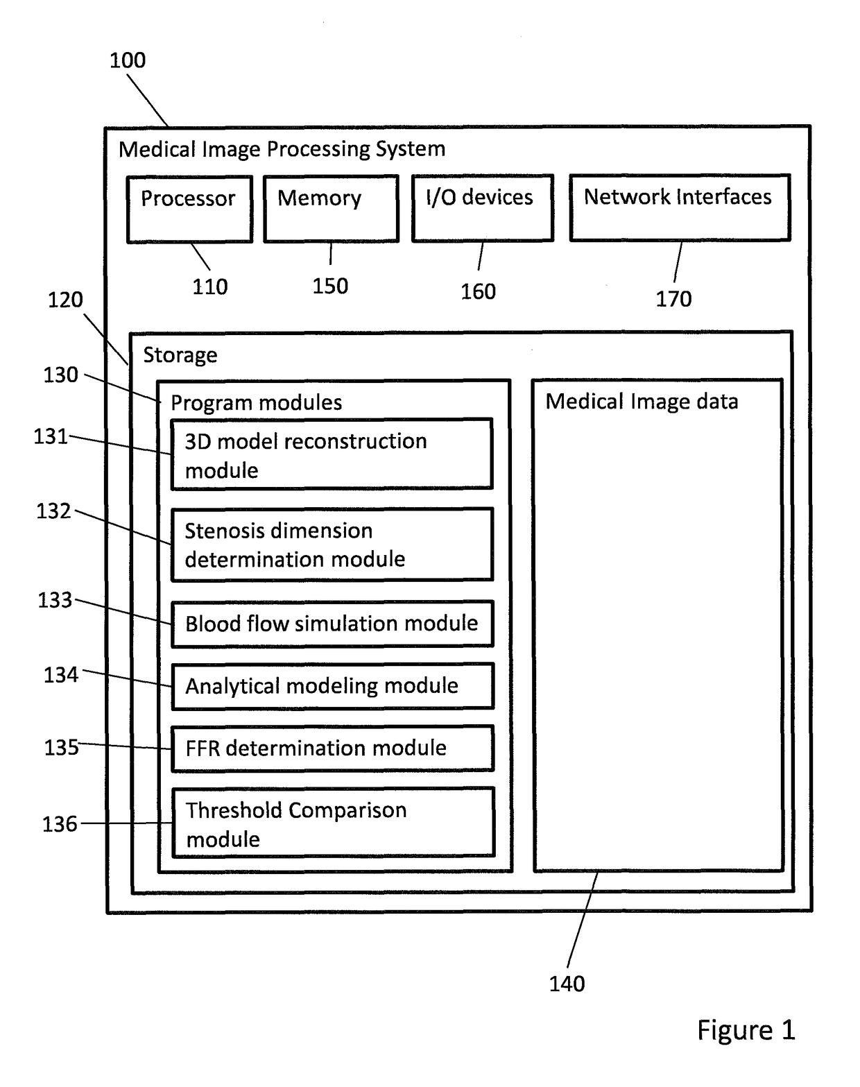
[0029]FIG. 1 shows a medical image processing system according to an embodiment of the present invention. The image processing system 100 comprises a processor 110 which may be referred to as a central processing unit (CPU) that is in communication with memory devices including storage 120 and memory 150. The Processor 110 may be implemented as one or more CPU chips. The memory 150 is implemented as a random access memory (RAM). The medical image processing system further comprises input / output (I / O) devices 160 and network interfaces 170.

[0030]The storage 120 typically comprises one or more disk drives and is used for non-volatile storage of data. The storage 120 stores program modules 130 which are loaded into the memory 150 when such programs are selected for execution. The program modules comprise a 3D model reconstruction module 131; a stenosis dimension determination module 132; a blood flow simulation module 133; an analytical modeling module 134; a fractional flow reserve (F...


PUM


Abstract

A medical image processing method of determining a fractional flow reserve through a stenosis of a coronary artery from medical image data is disclosed. The medical image data comprises a set of images of a coronary region of a patient. The coronary region includes the stenosis. The method comprises: reconstructing a three dimensional model of a coronary artery tree of the patient from the medical image data; determining stenosis dimensions from the three dimensional model of the coronary artery tree of the patient; simulating blood flow in the three dimensional model of the coronary artery tree of the patient to determine modeled flow rates; using an analytical model depending on the stenosis dimensions to predict a modeled pressure drop over the stenosis from the modeled flow rates; and determining the fractional flow reserve through the stenosis from the modeled pressure drop.

Description

FIELD OF THE INVENTION[0001]Embodiments of the present invention relate to medical image processing. More specifically, embodiments disclosed herein relate to the processing of medical images of a coronary region of a patient to assess coronary stenosis by determining a fractional flow reserve (FFR).BACKGROUND[0002]Cardiovascular disease is the leading cause of death in both developed and developing countries. It accounts for 30% of all global deaths. Due to the aging population in the world, the number of patients with cardiovascular disease is expected to dramatically increase. Coronary artery disease (CAD) is the most prevalent cardiovascular disease, caused by the build-up of plaque inside the coronary artery. The plaque narrows the artery and eventually impacts the blood supply to the heart. As a result, CAD has been linked to the development of cardiovascular-related events and accounts for 45% of deaths due to cardiovascular disease.[0003]Therefore characterization and quanti...

Claims


Application Information

Patent Timeline
IPC IPC(8): G06T7/00G06T11/00G06T7/60A61B6/00A61B6/03G06F19/00G16H30/20
CPCG06T7/0016G16H50/50G06T7/60A61B6/507A61B6/032A61B6/504A61B6/5217G06F19/321G06F19/3437G06T2207/30048G06T2207/30104G06T2200/08G06T2207/10081G06T2207/20076G06T11/003G06T2207/30096G06T19/20G06T2210/41A61B6/5205G06T7/0012G06T2207/10088G16H30/20
Inventor ZHONG, LIANGZHANG, JUN MEITAN, RU SAN
Owner SINGAPORE HEALTH SERVICES PTE
Why Eureka
Try Eureka