Devices, systems, and methods for fluorescence lifetime imaging microscopy

a lifetime imaging and microscopy technology, applied in the field of fluorescence lifetime imaging microscopy devices, systems and methods, can solve the problems of high cost and use of multiple embryos, high rates of multiple gestation, and low success rate of ar

Inactive Publication Date: 2018-05-03
PRESIDENT & FELLOWS OF HARVARD COLLEGE +1
View PDF0 Cites 4 Cited by
  • Summary
  • Abstract
  • Description
  • Claims
  • Application Information

AI Technical Summary

Benefits of technology

[0021]In all aspects of the invention, the methods are performed in vitro. The noninvasive nature of the methods allows them to be performed without harming the embryos or oocytes.

Problems solved by technology

However, success rates for ART are low, only 10%-35% of cycles result in successful birth, leading to high costs and use of multiple embryos, which in turn gives rise to high rates of multiple gestations.
Multiple gestations greatly increase mortality rates and suffering for both infant and mothers, and produce substantial financial costs.
One of the major reasons for the low success rate of ART is the absence of reliable methods for selecting the highest quality embryo(s) for transfer.
However, selection criteria generally remain subjective and only result in the ˜35% success rate quoted above.
Newly proposed non-microscopy based methods, using genomic, transcriptomic or proteomic based assays (Uyar, et al, 2012), require a biopsy of the embryo and are thus invasive and significantly reduce rates of embryo survival (Scott et al., 2006).
However, a prospective randomized trial has recently failed to show that utilization of such metabolomic assessment improves selection over morphologic evaluation alone (Vergouw et al, 2012).

Method used


Image

  • Devices, systems, and methods for fluorescence lifetime imaging microscopy

Examples


examples

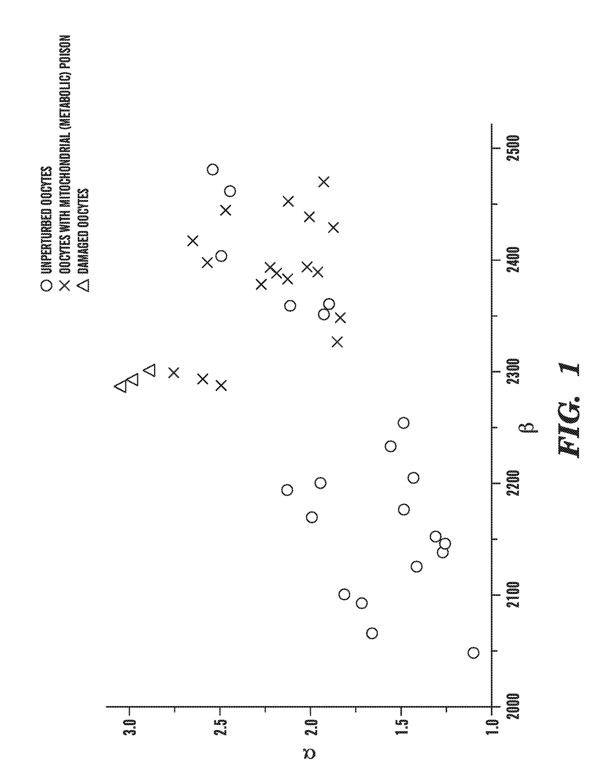
[0097]We obtained preliminary data on FLIM of NADH in mouse oocytes. The preliminary data was acquired on a FLIM system. The microscope consists of a ti-sapphire femtosecond laser (Spectra-Physics), an inverted microscope base (Nikon), a scan head (Becker & Hickl), a hybrid PMT detector (Hamamatsu), and electronics for time correlated single photon counting (Becker & Hickl). This microscope was assembled to acquire the preliminary data.

[0098]Oocytes were placed in a medium on the microscope stage and imaged. A single image of each oocyte was analyzed by averaging the FLIM data over the entire oocyte. The acquired fluorescence lifetime histogram from NADH, averaged over the entire oocyte, was fit to a sum of two exponentials. The parameter alpha, is the ratio of the amplitude of the two exponentials, the parameter, beta, is the lifetime of the longer exponential (in picoseconds).

[0099]Unperturbed oocytes exhibit a range of values of alpha and beta (FIG. 1, circles). As seen in FIG. 1...


PUM

PropertyMeasurementUnit
wavelengthaaaaaaaaaa

Abstract

The invention provides novel non-invasive in vitro methods for assessing the metabolic condition of oocytes and/or embryos with fluorescence lifetime imaging microscope, that can be used, for example, in assessment of oocytes and embryos in assisted reproductive technologies.

Description

CROSS-REFERENCE TO RELATED APPLICATION[0001]This application is a continuation of co-pending U.S. application Ser. No. 14 / 759,591 filed on Jul. 7, 2015 which is a 35 U.S.C. § 371 National Phase Entry Application of International Application No. PCT / US14 / 10435 filed Jan. 7, 2014, which claims benefit under 35 U.S.C. § 119(e) of U.S. Provisional Application No. 61 / 750,061 filed Jan. 8, 2013, the contents of which are incorporated herein by reference in their entireties.FIELD OF THE INVENTION[0002]The invention relates to methods for assessing the metabolic condition of oocytes and / or embryos, and can be used, e.g., in assessment of oocytes and embryos in assisted reproductive technologies.BACKGROUND[0003]Assisted Reproductive Technology (ART) has revolutionized the treatments of human infertility in the past 30 years and has become ubiquitous. ART currently accounts for over 1% of birth in the U.S. (SART, 2005). However, success rates for ART are low, only 10%-35% of cycles result in ...

Claims


Application Information

Patent Timeline
Patent Type & Authority Applications(United States)
IPC IPC(8): G01N21/64
CPCG01N21/6486G01N21/6408G01N2201/06193G01N21/6458A61B17/425G01N2201/12761G01N2021/6423G01N21/6428
Inventor RACOWSKY, CATHERINEDENG, MANQINEEDLEMAN, DANIEL
Owner PRESIDENT & FELLOWS OF HARVARD COLLEGE
Why Eureka
Try Eureka