Model-based segmentation of an anatomical structure

an anatomical structure and model technology, applied in the field of model-based segmentation of anatomical structures, can solve the problems of automatic segmentation algorithms that may at times produce erroneous segmentation and automatic algorithms may err in the selection of models, so as to achieve less time-consuming effects

Active Publication Date: 2018-05-17
KONINKLJIJKE PHILIPS NV
View PDF3 Cites 2 Cited by
  • Summary
  • Abstract
  • Description
  • Claims
  • Application Information

AI Technical Summary

Benefits of technology

[0032]Optionally, the medical image is a 3D medical image, the view is a 2D representation of the 3D medical image, and the plurality of models are 3D models. The user is thus e

Problems solved by technology

Automatic segmentation algorithms may at times produce erroneous segmentation results.
In particular, if different mo

Method used


Image

  • Model-based segmentation of an anatomical structure
  • Model-based segmentation of an anatomical structure
  • Model-based segmentation of an anatomical structure

Examples


Embodiment Construction

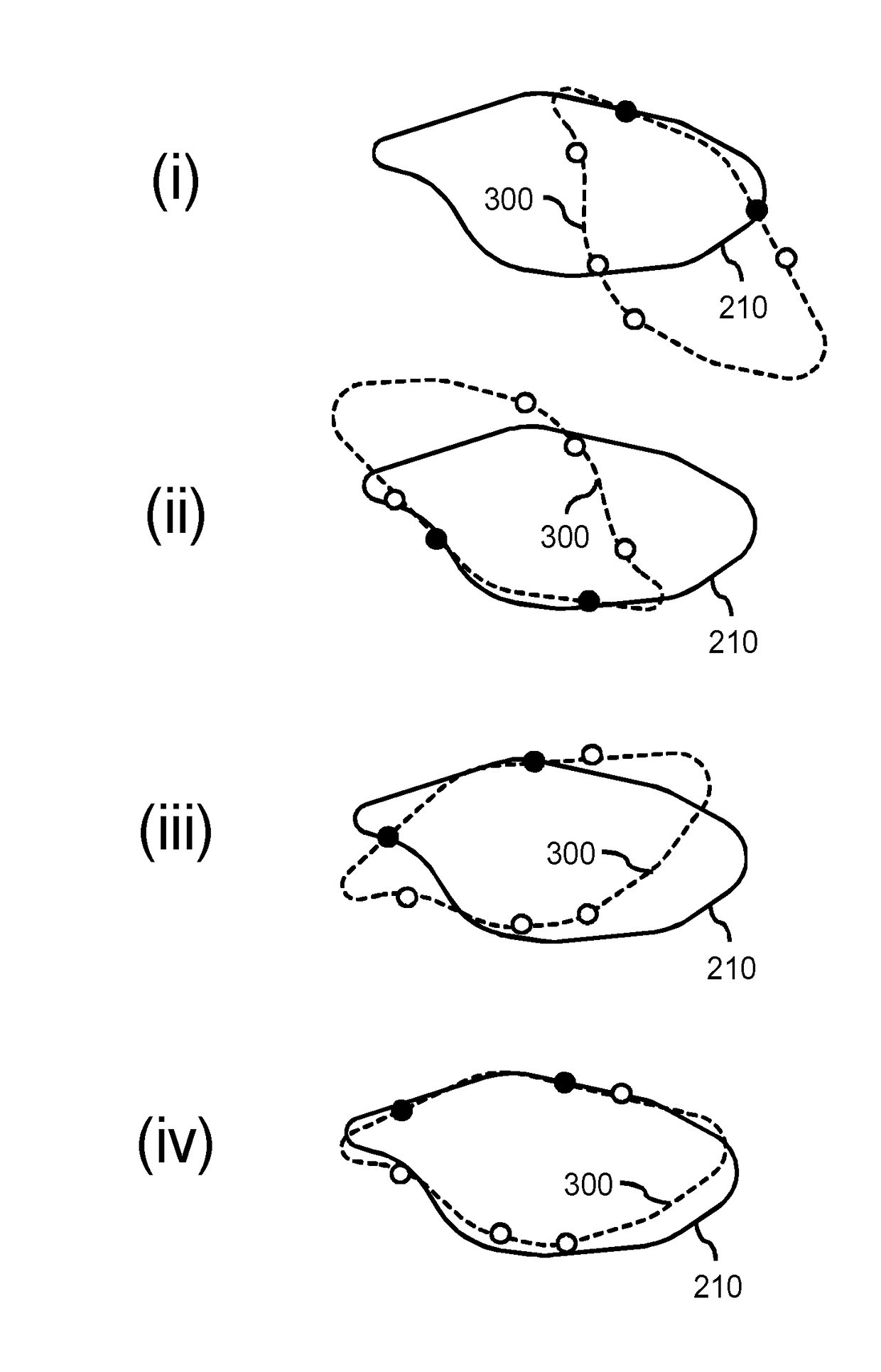
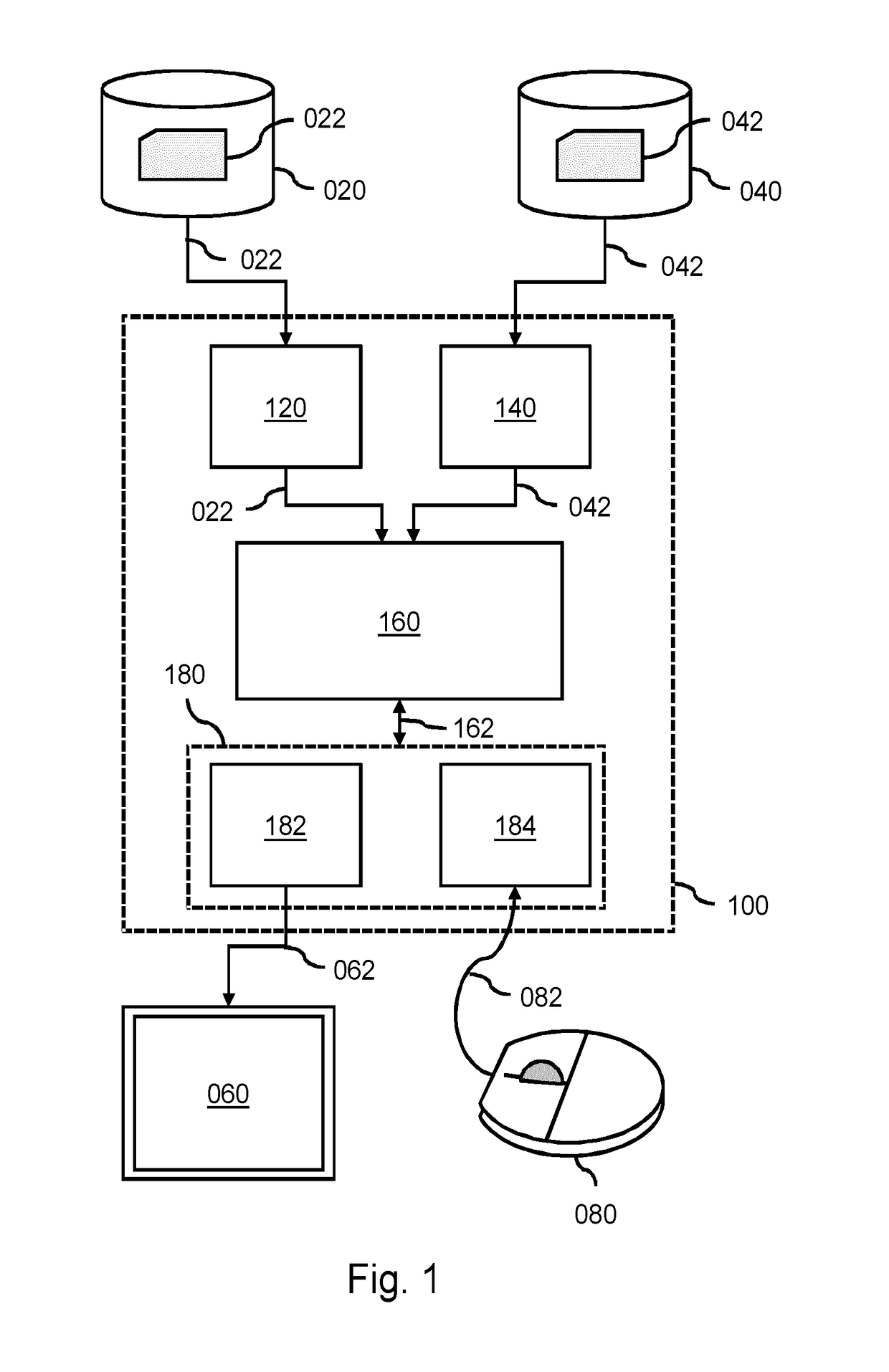
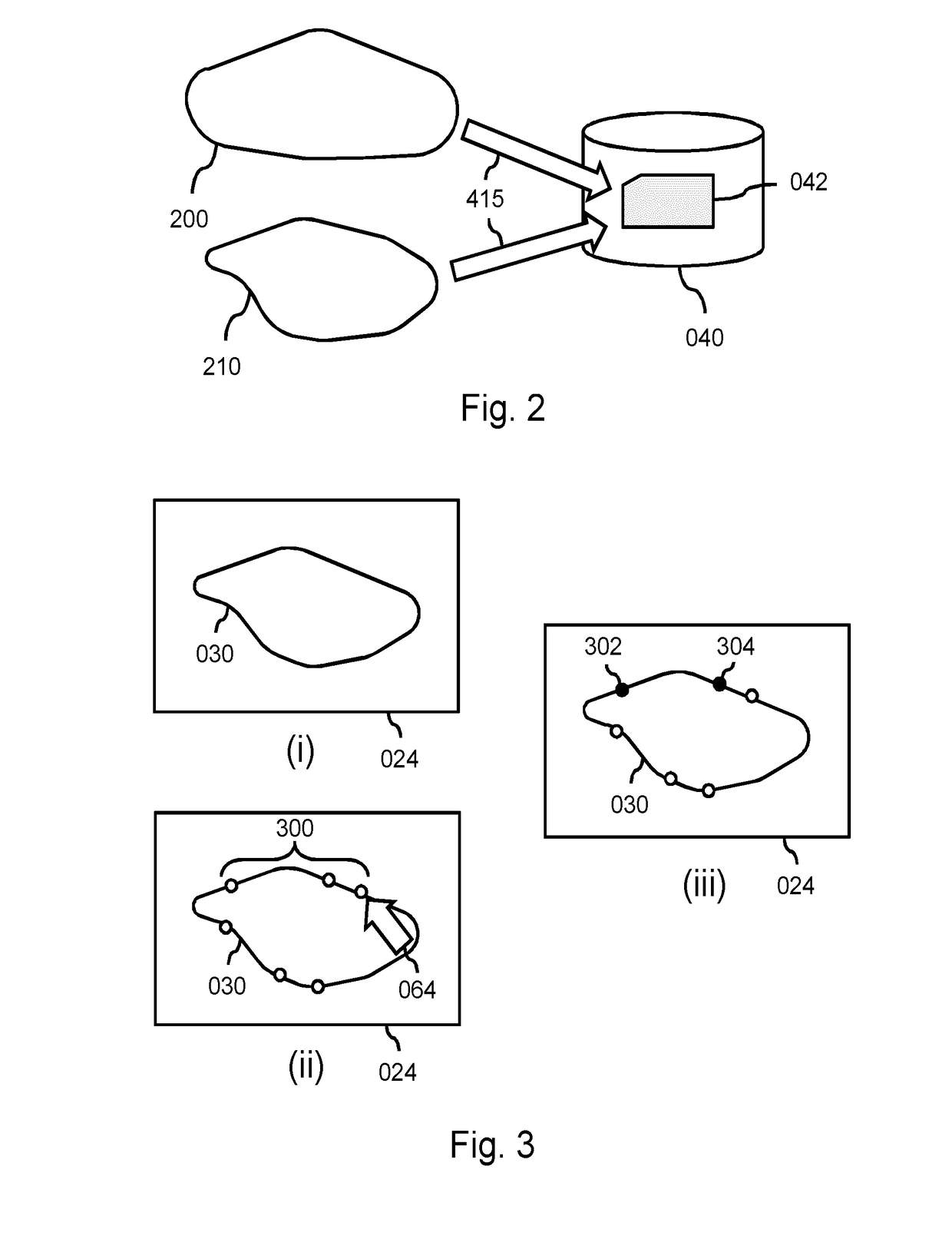
[0080]FIG. 1 shows a system 100 for segmentation of an anatomical structure in a medical image based on a limited set of user-indicated boundary points. Such a system may be employed in various medical applications, including, but not limited to, image annotation. The system 100 essentially involves a user interactively specifying the limited set of boundary points of the anatomical structure in a view of the medical image. The set of boundary points may, on its own, be considered an insufficient segmentation of the anatomical structure in the medical image, but is rather used to select a segmentation model from a plurality of different segmentation models. The selection is based on a goodness-of-fit measure between the boundary points and each of the segmentation models. For example, a best-fitting model may be selected and used for segmentation of the anatomical structure.

[0081]The system 100 comprises an image data interface 120 for accessing image data 022 of a medical image. Th...


PUM


Abstract

A system and method are provided for segmentation of an anatomical structure in which a user may interactively specify a limited set of boundary points of the anatomical structure in a view of a medical image. The set of boundary points may, on its own, be considered an insufficient segmentation of the anatomical structure in the medical image, but is rather used to select a segmentation model from a plurality of different segmentation models. The selection is based on a goodness-of-fit measure between the boundary points and each of the segmentation models. For example, a best-fitting model may be selected and used for segmentation of the anatomical structure. It is therefore not needed for the user to delineate the entire anatomical structure, which would be time consuming and ultimately error prone, nor is it needed for a segmentation algorithm to autonomously have to select a segmentation model, which may yield an erroneous selection.

Description

FIELD OF THE INVENTION[0001]The invention relates to a system and a method for performing a model-based segmentation of an anatomical structure in a medical image. The invention further relates to a workstation and imaging apparatus comprising the system and to a computer program product comprising instructions for causing a processor system to perform the method.BACKGROUND OF THE INVENTION[0002]Robust automatic segmentation of various anatomical structures in a medical image is a key enabler in improving clinical workflows. Here, the term segmentation refers to the identification of the anatomical structure in the medical image, e.g., by delineation of the boundaries of the anatomical structure, by labeling of the voxels enclosed by the boundaries, etc. Once such segmentation has been performed, it is possible to extract clinical parameters such as, in case of, e.g., a cardiac structure, ventricular mass, ejection fraction and wall thickness. When overlaid over, or otherwise applie...

Claims


Application Information

Patent Timeline
IPC IPC(8): G06T7/149G06T7/00G06T7/12G06K9/62
CPCG06T7/149G06T7/0012G06T7/12G06K9/6204G06K2209/051G06T2207/20101G06T2200/04G06T2210/41G06T2207/10072G06T2207/10116G06T2207/20096G06T2207/30048G06V10/752G06V2201/031G06V10/7788A61B5/00G06F18/41
Inventor LAWRENSON, MATTHEW JOHNNOLAN, JULIAN CHARLESWEESE, JUERGEN
Owner KONINKLJIJKE PHILIPS NV
Why Eureka
Try Eureka