

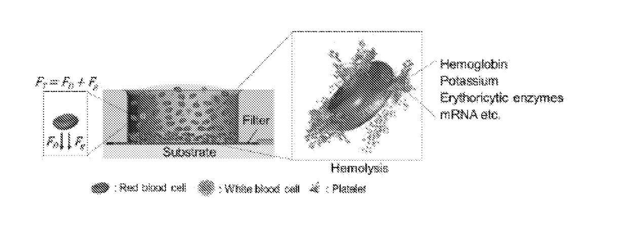
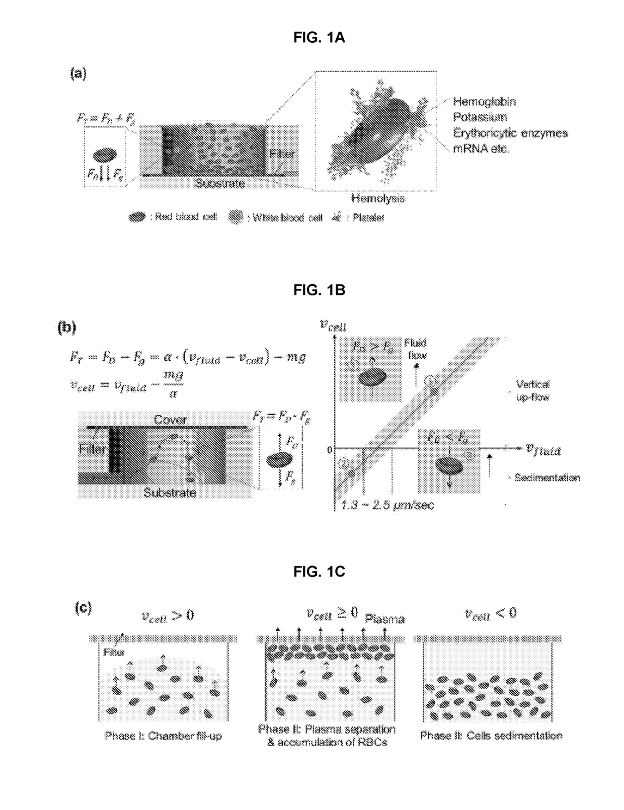
Hemolysis-free blood plasma separation
a technology of blood plasma and separation method, which is applied in the field of hemolysis-free blood plasma separation, can solve the problems of unsuitable poc application techniques, possible plasma contamination, and time-consuming and labor-intensive methods, and achieves the effects of reducing cell clogging, reducing clogging, and reducing the hemolysis of rbcs during separation
- Summary
- Abstract
- Description
- Claims
- Application Information
AI Technical Summary
Benefits of technology
Problems solved by technology
Method used
Image

Examples
example 1
Experimental
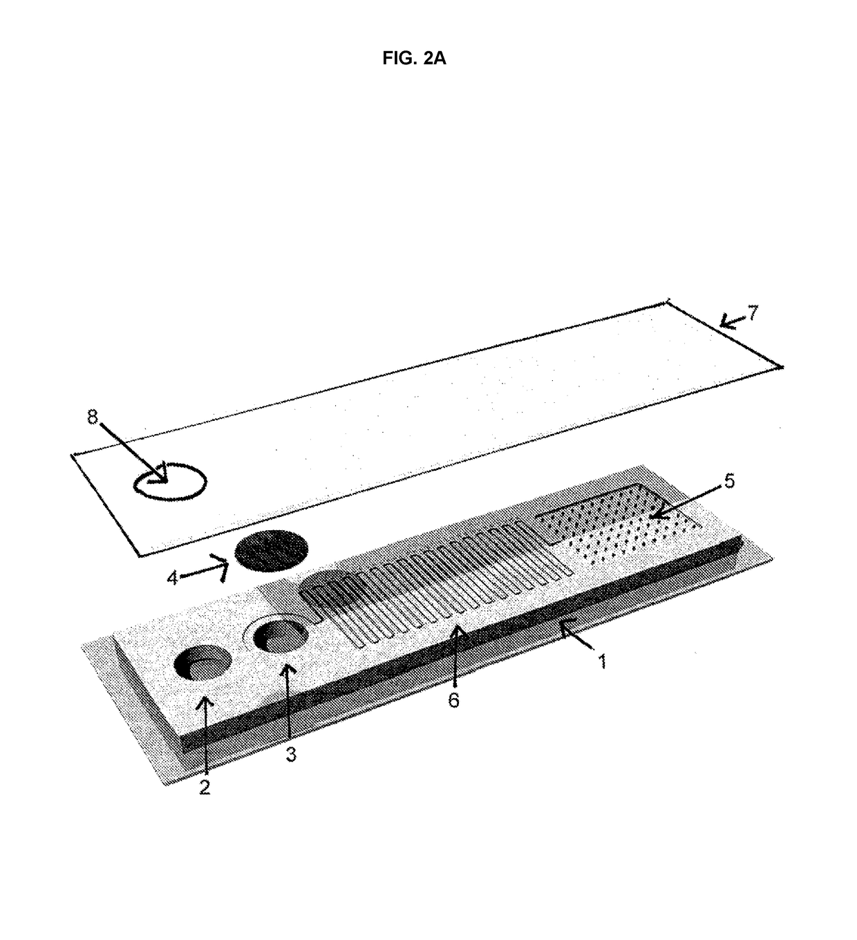
[0104]For the filter-in-top configuration, the device was fabricated by placing a 1.8 mm thick poly(dimethylsiloxane) (PDMS) layer between two 1 mm thick plastic sheets (PSS-3000, Piedmont Plastics) (FIG. 2I and FIG. 2J). The PDMS layer consisted of two separate PDMS layers, bonded together after O2 plasma treatment. The top and bottom microfluidic channels face the top and bottom plastic sheets, respectively. Top channels contain a ruler to measure the volume of separated plasma. The two channels were connected by a vertical up-flow channel created with a variable size of Harris Uni-Core™ punch. A Nuclepore™ track-etched polycarbonate membrane filter with 0.4 μm pores purchased from Whatman was used for blood plasma separation. The membrane filter was placed between the top PDMS layer and plastic sheet. For the filter-in-bottom device, a 1.8 mm thick single PDMS layer with ruler was bonded with a bottom plastic sheet. The filter was placed below the inlet of the device....
PUM
 Abstract
Description
Claims
Application Information