Protein therapy for treatment of retinal diseases

a technology for retinal diseases and proteins, applied in the field of protein therapy for the treatment of retinal diseases, can solve the problems of limited or no benefit in therapy, increased number of dr patients, and significant morbidity in the u.s. and throughout the world, and achieve the effect of facilitating the expression of polypeptides and facilitating the transfer of fusion proteins

Inactive Publication Date: 2018-08-23
SCOTT & WHITE HEALTHCARE
View PDF1 Cites 9 Cited by
  • Summary
  • Abstract
  • Description
  • Claims
  • Application Information

AI Technical Summary

Benefits of technology

[0020]The present invention also includes methods of treating a subject with retinal degeneration comprising administering to a subject with retinal degeneration a pharmaceutically effective amount of a composition comprising mesenchymal stem cells, wherein the injection is intravitreal or subretinal. In particular embodiments, the retinal degeneration is retinitis pigmentosa or age-related macular degeneration. In some embodiments, the stem cells have been genetically modified to overexpress a stanniocalcin family member polypeptide as set forth herein. In a particular embodiment, the stanniocalcin family member polypeptide is an STC-1 polypeptide. Overexpression of the stanniocalcin family member polypeptide is overexpression compared to the expression of the same stanniocalcin family member polypeptide by mesenchymal stem cells that have not been genetically modified.
[0021]Also included as part of the invention are kits that include (a) a pharmaceutically effective amount a stanniocalcin family member polypeptide or a TSG-6 family member polypeptide in one or more sealed vials. The stanniocalcin family member polypeptide and TSG-6 polypeptide may include any of the sequences previously set forth. The polypeptide may have between 95% and 99% sequence identity to any of the foregoing sequences. In a particular aspect, the kit includes a pharmaceutically effective amount of a STC-1 polypeptide that has at least 95% sequence identity to SEQ ID NO:1. In further aspects, the kit includes a syringe. For example, the syringe may be a tuberculin syringe. The kit may include a 25-gauge needle or a 30-gauge needle. The kit may optionally include instructions for use of the polypeptide either written on a paper or in a computer-readable format. The kit may optionally further include a 1 cc syringe or a 2 cc syringe. In a particular aspects, the stanniocalcin family member polypeptide comprises SEQ ID NO:1. In a particular aspect, the polypeptide is in a lyophilized form, and the kit further includes instructions for reconstituting the lyophilized polypeptide into a carrier for administration to a subject. For example, the carrier may be sterile water, normal saline, or phosphate buffered saline. The carrier may be comprised in one or more separate vials. In some aspects, the kit includes a polypeptide as set forth herein comprised in an ophthalmic drug delivery device, including a biodegradable drug-eluting device.
[0022]Other embodiments of the present invention concern methods of treating an eye disease comprising administering to a subject a pharmaceutically effective amount of a polynucleotide expressing a STC-1, STC-2, or TSG-6 polypeptide. The STC-1, STC-2, or TSG-6 polypeptide may be selected from any of the aforementioned polypeptide sequences. In some aspects, the polynucleotide is comprised in an expression cassette wherein the polynucleotide is operatively coupled to a promoter that facilitates expression of the polypeptide in a target cell. In some aspects, the expression cassette is comprised in a vector, such as a cell (such as a MSC), viral vector, a liposome, or a nanoparticle.
[0023]Fusion proteins and polynucleotides encoding them (RNA, and DNA, such as DNA polynucleotides in expression vectors) are also contemplated as part of the invention. A fusion protein is a single polypeptide sequence created through the joining of two or more genes which originally coded for separate polypeptides, with functional properties derived from the original polypeptides. The fusion proteins include a first domain that includes a stanniocalcin family member polypeptide or a TSG-6 family member polypeptide and a second domain comprising a second therapeutic polypeptide or a carrier polypeptide to facilitate transfer of the fusion protein into a cell. In a particular aspect, the second therapeutic polypeptide is a CD59 polypeptide or an antiangiogenic polypeptide. The sequence of human CD59 is provided in SEQ ID NO:26 and is associated with GenBank accession number CAG46523. Nonlimiting examples of angiostatic polypeptides include ranibizumab, endostatin, bevacizumab, or aflibercept. Additional non-limiting examples of agents which may be present in the fusion protein or in other covalent or non-covalently associated complexes with a stanniocalcin family member polypeptide or a TSG-6 family member polypeptide are Lucentis, Macugen, Pegaptanib, Ranibizumab, Eylea, Verteporfin, Visudyne, an angiostatic cortisene formulation, such as anecortave acetate suspension (RETAANE). Nonlimiting examples of carrier polypeptides include poly-Arg, a Tat-derived amino acid sequence and Drosophila Antennapedia homeodomains. Numerous examples of carrier polypeptides are set for in U.S. Pat. No. 7,939,493, which is herein specifically incorporated by reference in its entirety. In this patent they are designated as “carrier peptides.” A “peptide” for purposes of the present patent application is an example of a “polypeptide” and the terms are used interchangeably herein. Other embodiments include a polynucleotide encoding a fusion protein of the present invention.
[0024]Ophthalmic drug delivery devices for intravitreal or subconjunctival delivery of the stanniocalcin family member polypeptide, a TSG-6 polypeptide, a fusion protein, or a stimulated or genetically modified cell as set forth herein are also contemplated as part of the present invention. The ophthalmic drug delivery device may comprise an effective amount of any of the foregoing polypeptides, fusion proteins, and cells set forth herein. In some embodiments, the polypeptide has between 95% and 99% sequence identity to any of the foregoing sequences. The polypeptide may be enclosed in a reservoir or in contact with a surface of the drug delivery device. In some aspects, the drug delivery device is a scleral-fixated nonbiodegradable implant. In other embodiments, the drug delivery device is a biodegradable implant designed to be free-floating in the vitreous cavity. In some embodiments, the drug delivery device further comprises cells that express the STC-1 polypeptide in the reservoir or in contact with a surface of the drug delivery device. In particular aspects, the ophthalmic drug delivery device is OZURDEX™, VITRASERT™, I-VATION™, RETISERT™, or ILUVIEN™. Other non-limiting examples of drug delivery devices include Ocusert®, collagen shields, or a delivery device comprising polyacrylic acid, polyvinyl alcohol, silicone elastomer, hydroxy propyl cellulose, ethyl cellulose, cellulose acetate phthalate and polymethacrylic acid, or hyaluronic acid.
[0025]Also included are pharmaceutical compositions for ophthalmic delivery comprising a therapeutically effective amount of at least one therapeutic polypeptide selected from any of Tables 1, 2, or 3, or a polypeptide that has at least 95% sequence identity to a protein set forth in any of Tables 1, 2, or 3 or a polypeptide that has between 95% and 99% sequence identity to any of the polypeptides set forth in Tables 1, 2, or 3. The pharmaceutical composition may optionally comprise one or more additional therapeutic agents suitable for ophthalmic delivery. Non-limiting examples of additional therapeutic agents include antibiotics, anti-inflammatory agents, anti-proliferative agents, anti-neovascular agents (such as agents which antagonize the function of neovascular growth factors (i.e., vascular endothelial growth factor (VEGF), endothelial cell surface receptors, and / or extracellular matrix (ECM) proteins, which are important mediators of neovascularization), anti-viral agents (i.e., idoxuridine, vidarabine, trifluorothymidine), beta-andrenergic blockers (timolol maleate, levobunolol), corticosteroids, retinoic acid formulations, vitamins, topical anesthetics (i.e., proparacaine hydrochloride, tetracaine hydrochloride), and the like. Other examples include bevacizumab (Avastin), pegaptanib (Macugen), Lucentis (ranibizumab), verteporfin (Visudyne), and CD59.

Problems solved by technology

Eye disease is a significant cause of morbidity in the U.S. and throughout the world.
While therapies have improved over time for many eye diseases, there remain many others for which therapy is of limited or no benefit.
Despite adequate glycemic and blood pressure control and lipid-lowering therapy, the number of DR patients continues to grow and therapeutic approaches remain limited.
Retinal vessel occlusion and degeneration is a typical feature of DR and is also a cause of neovascularization.
Dry, atrophic (nonexudative) age-related macular degeneration, defined as progressive age-related degeneration of the macula associated with retinal pigment epithelial changes including atrophy and drusen, is a common cause of vision loss in adults for which therapy is extremely limited.
Vitamin therapies and other types of therapy are of limited benefit.
Nevertheless, despite therapy such as laser or pharmacotherapy, many patients develop progressive vision loss.

Method used


Image

  • Protein therapy for treatment of retinal diseases
  • Protein therapy for treatment of retinal diseases
  • Protein therapy for treatment of retinal diseases

Examples


example 1

MSC-Derived Factors Suppressed Inflammation and Neovascularization, and Promoted Wound Healing in Chemically-Injured Rat Cornea

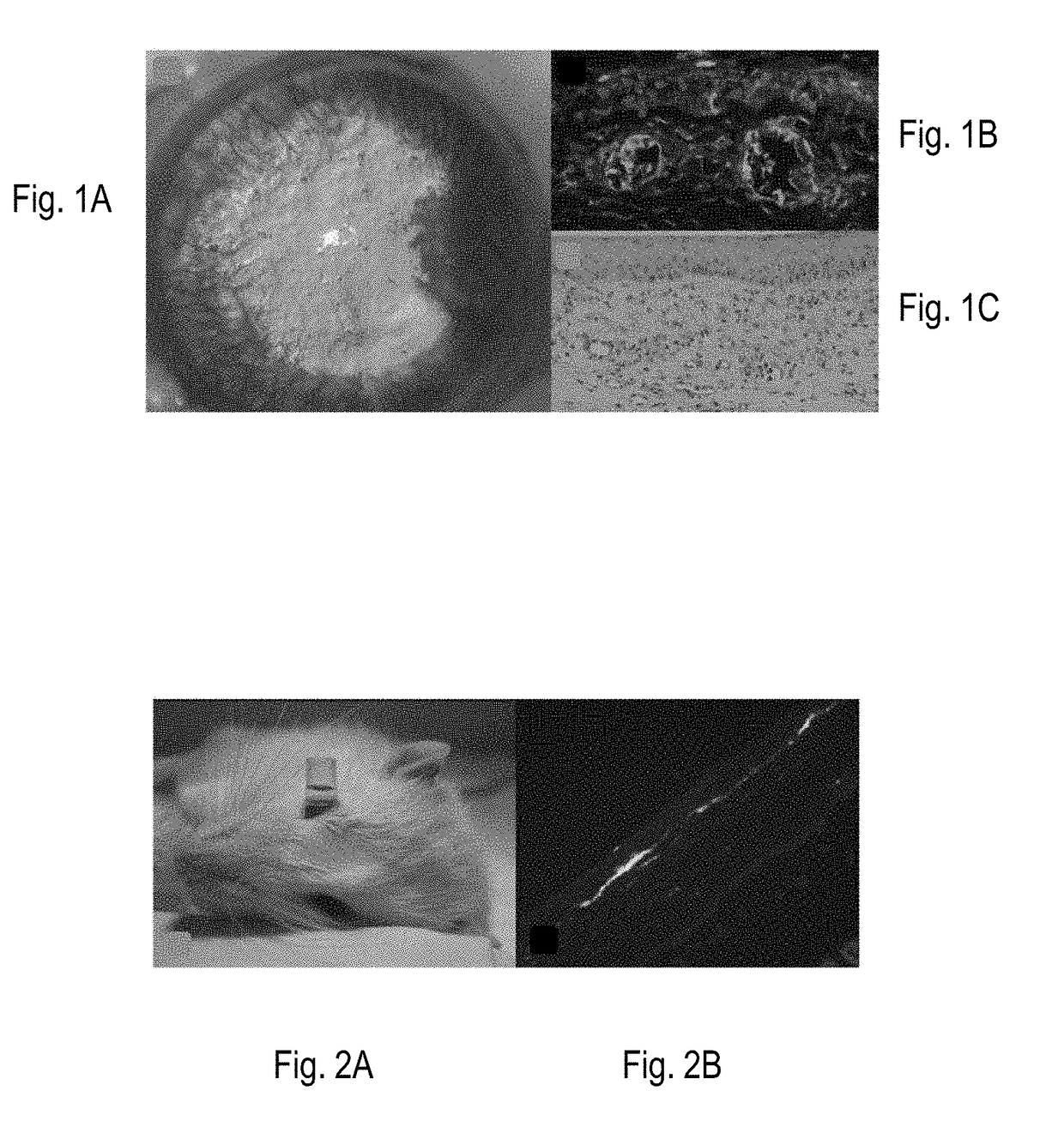
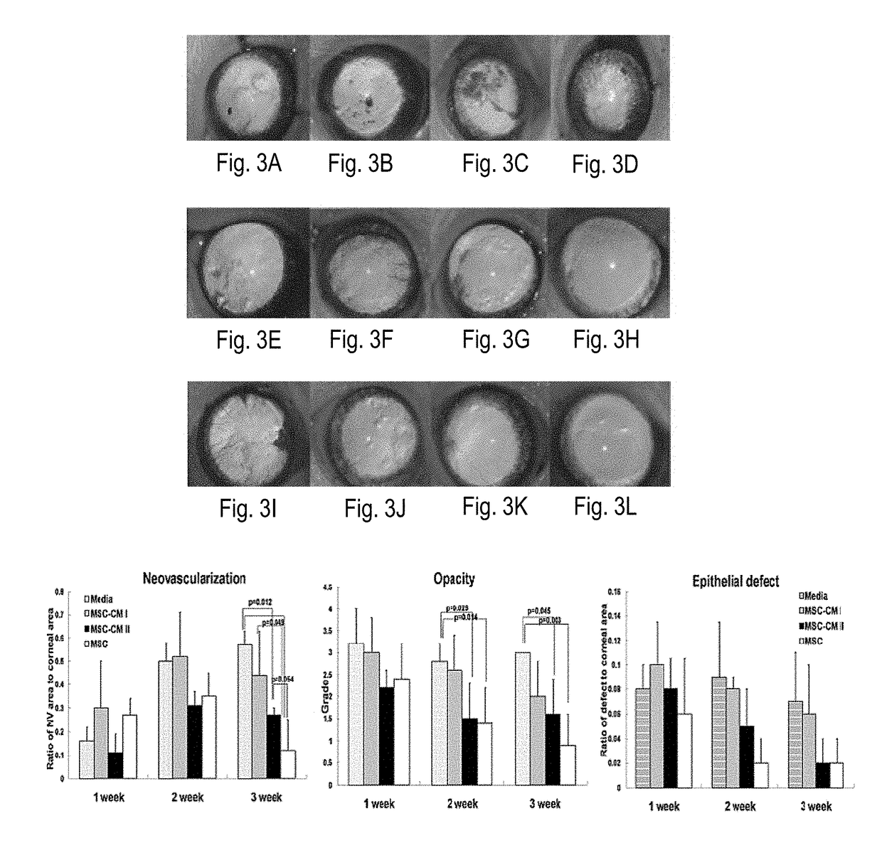
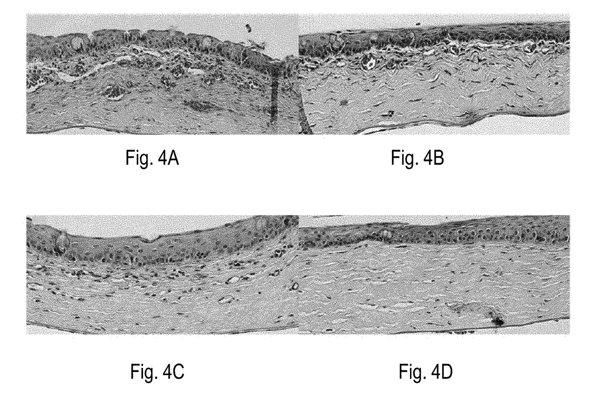
[0230]Beneficial effects of MSCs and MSC-derived factors have been observed in suppressing corneal inflammation / neovascularization and promoting wound healing (Oh et al., 2008). Corneal inflammation, neovascularization, and delayed wound healing were induced in rats by applying 100% ethanol for 30 sec and scraping both the epithelium in the limbus and the whole cornea. The reliability and reproducibility of this model was previously confirmed and repetitively used by other researchers (Cho et al., 1998; Avila et al., 2001; Ti et al., 2002; Espana et al., 2003; Homma et al, 2004; Oh et al., 2009b). Massive infiltration of inflammatory cells and growth of new vessels in cornea were induced in this model (FIG. 1).

[0231]Immediately after injury, rat MSCs or conditioned media (CM) derived from MSC cultures were put into an applicator and allowed to remain in the ...

example 2

-Conditioned Media Rescued Human Corneal Epithelial Cells from Chemically-Induced Apoptosis

[0236]Human corneal epithelial cells (hCECs) were chemically damaged by incubation in 15% ethanol for 30 seconds. Damaged hCECs were cultured with one of the following: (1) hMSC-conditioned media, (2) conditioned media from hMSC-damaged hCECs coculture, or (3) fresh media. Then, survival of hCECs was evaluated with MTT assay. The result showed that the proportion of damaged hCECs was significantly decreased when cultured with hMSC-conditioned media (FIG. 7).

example 3

n of MMP-9 is Significantly Suppressed in Chemically-Damaged Human Corneal Epithelial Cells by hMSCs

[0237]The following experiments were performed to evaluate how MSCs affected corneal epithelial cells in terms of inflammatory and angiogenic cytokine secretion. The hCECs were chemically damaged, then they were cocultured with hMSCs for 24 hours, and finally the cell-free supernatant was analyzed for cytokine concentration by ELISA. The coculture groups were as follows: (I) hPBMCs (human peripheral blood mononuclear cells), (2) hCECs, (3) hPBMCs / hCECs, (4) hMSCs, (5) hMSCs / hPBMCs, (6) hMSCs / hCECs, and (7) hMSCs / hPBMCs / hCECs. As a result, it was observed that MSCs constitutively secreted VEGF, MMP-2, and TSP-1 (FIG. 8). It is important to note that MMP-9, which is highly secreted by damaged hCECs, was significantly suppressed by hMSCs (FIG. 8, upper right). In fact, as a consequence of hMSC suppression the level of MMP-9 was reduced from 100% to 8%. Based on these results, it was beli...


PUM

PropertyMeasurementUnit
Sizeaaaaaaaaaa
Sizeaaaaaaaaaa
length measurementaaaaaaaaaa

Abstract

The present invention encompasses methods, compositions, and devices for treating an ocular disease, disorder or condition in a mammal. The invention includes polypeptides that possess anti-inflammatory, anti-apoptotic, immune modulatory and anti-tumorigenic properties, and their application in the treatment of eye disease, particularly diseases of the retina. In particular aspects, the invention includes administration of a therapeutic polypeptide such as a stanniocalcin family member protein for the treatment of an eye disease. Also included are fusion proteins and cells stimulated or modified to express the therapeutic polypeptides as set forth herein.

Description

CROSS-REFERENCE TO RELATED APPLICATIONS[0001]This patent application is a continuation-in-part of U.S. patent application Ser. No. 15 / 346,829, filed Nov. 9, 2016, which is a continuation of U.S. patent application Ser. No. 14 / 789,389, filed Jul. 1, 2015, now U.S. Pat. No. 9,498,517, which is a continuation of U.S. patent application Ser. No. 14 / 284,911, filed May 22, 2014, now U.S. Pat. No. 9,090,704, which is a continuation of U.S. patent application Ser. No. 13 / 549,770, filed Jul. 16, 2012, now U.S. Pat. No. 8,759,298, which claims priority to U.S. Provisional Patent Application No. 61 / 508,587, filed Jul. 15, 2011, and is a continuation-in-part application of International Patent Application No. PCT / US2011 / 000771, filed May 3, 2011, which in turn claims priority to U.S. Provisional Patent Application No. 61 / 464,172, filed Feb. 28, 2011, and to U.S. Provisional Patent Application No. 61 / 330,735, filed May 3, 2010, the contents of each of which of the foregoing applications are inco...

Claims


Application Information

Patent Timeline
Patent Type & Authority Applications(United States)
IPC IPC(8): A61K38/22A61K9/00C07K14/575A61K38/17A61K35/28
CPCA61K9/0048C07K14/575A61K38/1709A61K38/22A61K35/28
Inventor ROSA, ROBERT
Owner SCOTT & WHITE HEALTHCARE
Why Eureka
Try Eureka