Multi-layer airway organoids and methods of making and using the same

a multi-layer airway and organoid technology, applied in the field of multi-layer airway organoids, can solve the problems of inability to create the differentiated tissue components and structural complexity of the airway epithelium of most in vitro models, and inability to study respiratory infections in a significant way

Active Publication Date: 2018-10-11
WAKE FOREST UNIV HEALTH SCI INC
View PDF0 Cites 10 Cited by
  • Summary
  • Abstract
  • Description
  • Claims
  • Application Information

AI Technical Summary

Problems solved by technology

The study of respiratory infection is significantly limited by a lack of suitable in vivo and in vitro models to investigate interactions between the respiratory epithelium, infection and disease.
Additionally, most in vitro models are unable to create the differentiated tissue components and structural complexity of the airway epithelium.
However, these techniques present cells with artificial conditions, including two-dimensional (2D) growth surfaces that are several magnitudes stiffer than most soft tissues, and lack of important sig

Method used


Image

  • Multi-layer airway organoids and methods of making and using the same
  • Multi-layer airway organoids and methods of making and using the same
  • Multi-layer airway organoids and methods of making and using the same

Examples


examples

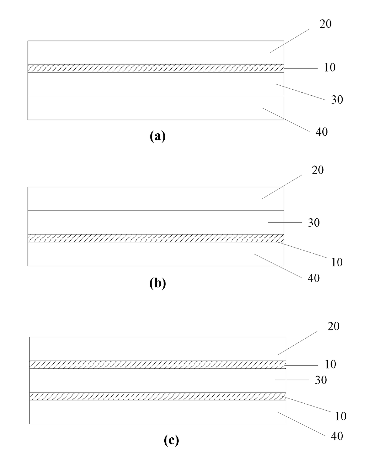
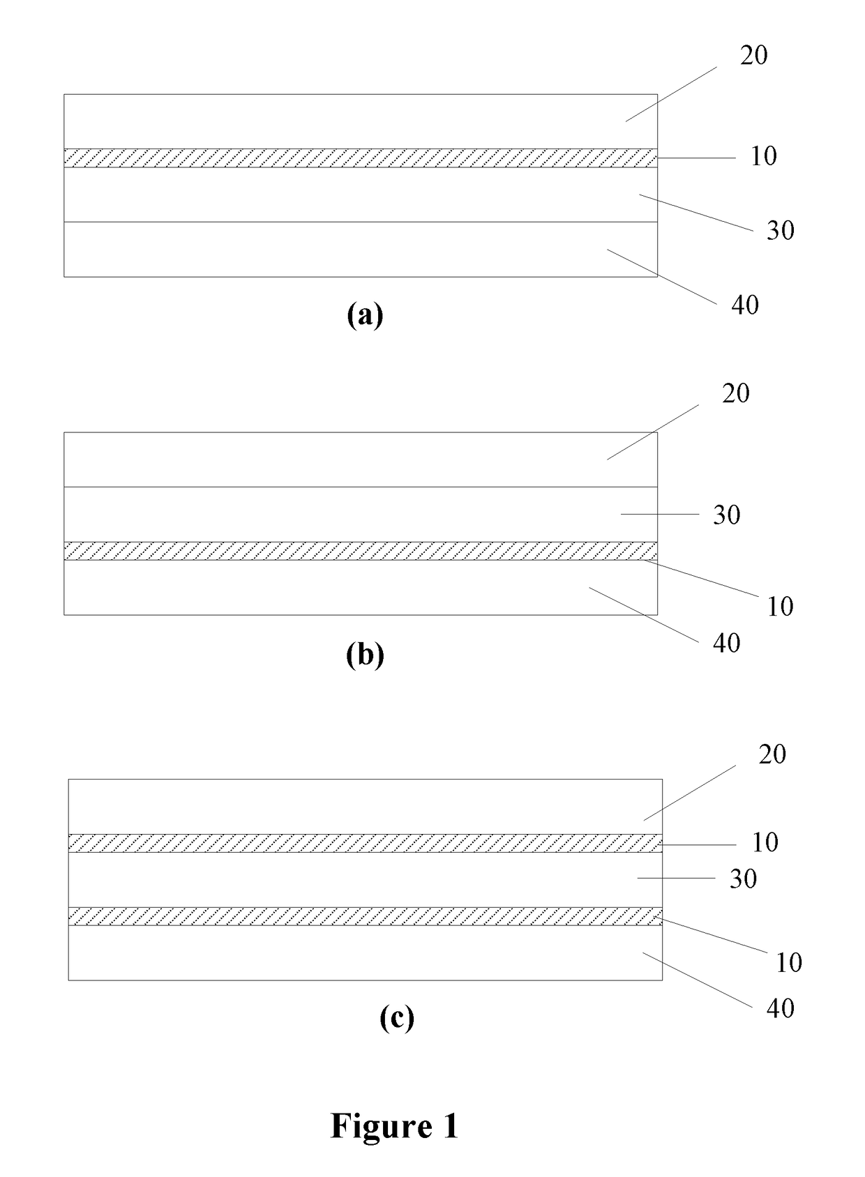
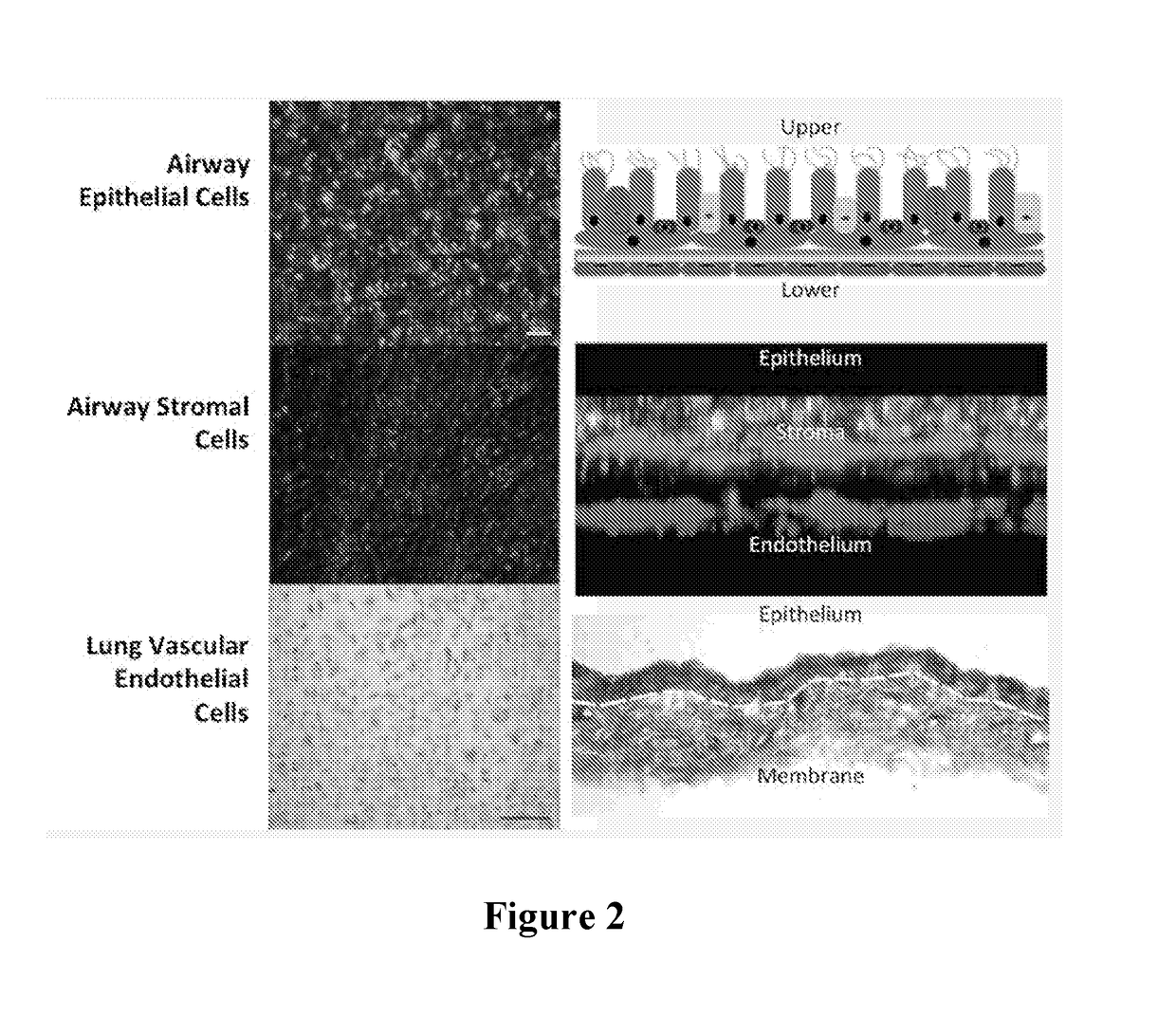
[0079]An airway organoid was constructed using primary normal human bronchial epithelial (NHBE) cells, primary human lung fibroblasts and human endothelial cells (HUVEC), as found in the normal human upper airway, layered on a polyester membrane in an order and ratio to replicate human airway tissue.

1. Construction of Lung Organoid

[0080]Each side of a polyester membrane (Corning® Costar® Snapwell™ cell culture inserts, 12 mm with 0.4 μm pore, pore density 4×10⁶ pores / cm2, polyester membrane, TC-treated, sterile) was coated with 150 μl collagen IV (Sigma C7521) and left under the biosafety hood overnight (hood open and blower on). The membrane was UV sterilized for 30 mins the next morning.

[0081]250,000 HUVECs (endothelial cells) (Cell and Viral Vector Core Laboratory, Wake Forest University) were seeded on the underside of the membrane and let stand for up to 4 hours for cells to attach. Membrane was then placed with 2 ml of EGM-10 in the well of the 6 well plate.

[0082]250,000 HALF ...


PUM


Abstract

Provided herein are artificial lung organoids. The artificial lung organoids may include an epithelial cell layer comprising mammalian lung epithelial cells, a stromal cell layer comprising mammalian lung fibroblast cells and an endothelial cell layer comprising mammalian endothelial cells. The artificial lung organoids may optionally include a porous membrane between said epithelial cell layer and said stromal cell layer and/or between said stromal cell layer and said endothelial lung cell layer.

Description

RELATED APPLICATIONS[0001]This application claims the benefit of priority from U.S. Provisional Patent Application Ser. Nos. 62 / 242,611, filed Oct. 16, 2015, and 62 / 404,931, filed Oct. 6, 2016, the contents of which are incorporated herein by reference in their entireties.FIELD OF THE INVENTION[0002]This invention relates generally to multi-layer airway organoids and methods of making and using of the same.BACKGROUND OF THE INVENTION[0003]The study of respiratory infection is significantly limited by a lack of suitable in vivo and in vitro models to investigate interactions between the respiratory epithelium, infection and disease. For example, animal models often do not acquire the pathological abnormalities in the airways and lungs seen in humans. See, e.g., Guilbault et al., Cystic fibrosis mouse models, American Journal of Respiratory Cell and Molecular Biology. 2007; 36(1):1-7. Additionally, most in vitro models are unable to create the differentiated tissue components and stru...

Claims


Application Information

Patent Timeline
IPC IPC(8): C12N5/071C12M3/00C12M3/06C12M1/00C12N1/20
CPCC12N5/0697C12M21/08C12M23/16C12M23/22C12N5/0688C12N1/20C12N2502/27C12N2502/28C12N2533/54C12N2533/90G09B23/28
Inventor MURPHY, SEAN V.ATALA, ANTHONY
Owner WAKE FOREST UNIV HEALTH SCI INC
Why Eureka
Try Eureka