Three dimensional vaginal tissue model containing immune cells

a vaginal tissue and three-dimensional technology, applied in the field of three-dimensional vaginal tissue models containing immune cells, can solve the problems of severe limitations in the use of vaginal tissue models, the lack of differentiated function and barrier properties of cells in monolayer culture, and the inability to achieve differentiation and barrier properties of normal cells, etc., to achieve the effect of increasing viability, promoting proliferation of immune cells, and increasing viability

Inactive Publication Date: 2005-09-13
MATTEK CORP
View PDF25 Cites 52 Cited by
  • Summary
  • Abstract
  • Description
  • Claims
  • Application Information

AI Technical Summary

Benefits of technology

[0018]In a preferred embodiment of the method, the seeding step is at a ratio of about 1:1 vaginal epithelial cells to immune cells. In another embodiment, the seeding step of the method is at a ratio of between about 1:1 and 10,000:1 vaginal epithelial cells to immune cells, and the co-culturing step is in serum-free medium supplemented w

Problems solved by technology

Although useful, each model system developed to date has suffered from significant drawbacks.
However, cells in monolayer culture lack the differentiated function and barrier properties of cells found in normal tissue.
Although useful, due to the skin origin of the cells, the tissue is not fully representative of in vivo cervical and vaginal tissue, and thus its use as a vaginal tissue model is severely limited.
However, the availability o

Method used


Image

  • Three dimensional vaginal tissue model containing immune cells
  • Three dimensional vaginal tissue model containing immune cells

Examples


example 1

Generation and Characterization of the Cervico-Vaginal Tissue Equivalent

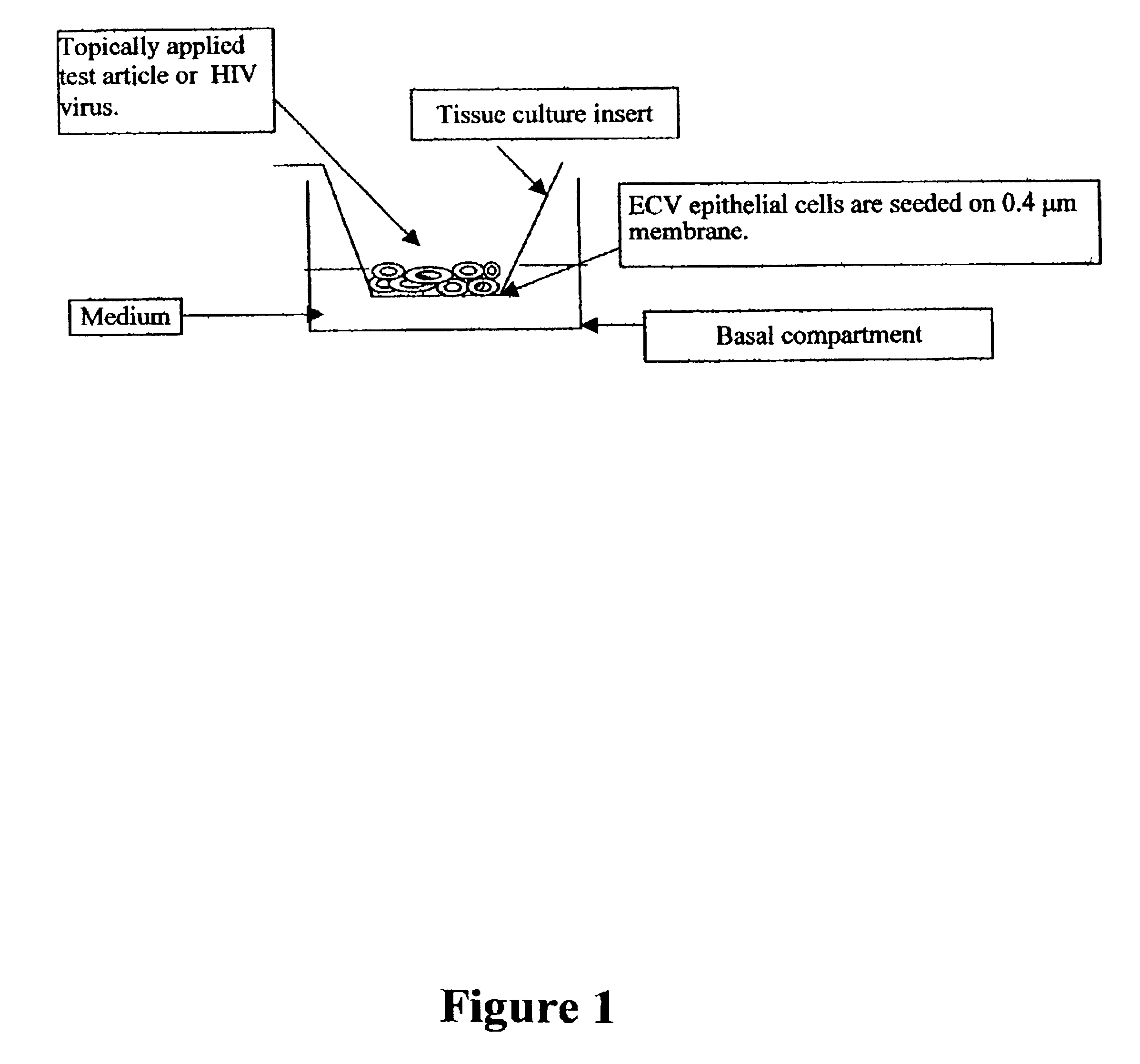
[0094]A source of both ecto-cervical, endo-cervico, or vaginal epithelial cells along with immune cells such as dendritic, Langerhans, or precursor cells, are necessary to produce the cervico-vaginal tissue equivalent. In this example, primary cervical epithelial cells were obtained from human ecto-cervical tissue. Langerhans cells (CD1a+ cells) were obtained by differentiating Langerhans precursor cells (CD34+ cells) using cytokines. Langerhans precursor cells were harvested from umbilical cord blood samples.

Epithelial Cells

[0095]Epithelial cells were obtained from ectocervical tissue and vaginal tissue obtained from women undergoing hysterectomies. After surgical removal of the tissue, the tissue was placed in Dulbecco's Modified Eagle's Medium (DMEM) containing 10× concentrate of penicillin streptomycin (1 mg / ml) and gentamicin (0.5 mg / ml) and stored at 4° C. until the tissue could be processed (within 24 hou...

example 2

Use of Langerhans Cells Derived from Monocytes to Generate the Cervico-Vaginal Tissue Equivalent

[0108]Langerhans cells isolated from monocytes performed equally well in the generation of the in vitro ectocervico-vaginal tissue. Experiments identical to those performed in Example 1 were performed on in vitro generated cervico-vaginal tissue equivalent which contained Langerhans cells generated from monocytes.

Generation of Langerhans Cells from Precursor Cells

[0109]Purified macrophages were negatively isolated from cord blood / adult blood derived mononuclear cells (MNC) with immuno-magnetic beads (Dynal Inc., Lake Success, N.Y.) following the manufacturer's recommendation, or by plastic adhesion. MNC (1×10⁷) were re-suspended in 200 μl PBS supplemented with 0.1% bovine serum albumin (BSA), 20 μl of blocking reagent, and 20 μl antibody mix (monocyte-kit provided by Dynal) and incubated at 4° C. for 10 min. Cells were then washed 2× and re-suspended in PBS containing 0.1% BSA. Depletion ...

example 3

Use of Langerhans Precursor Cells

[0111]This example is identical to Example 1 except that Langerhans precursor cells were used instead of Langerhans cells. Cytokines were added to the proliferation and differentiation medium to allow maturation of the Langerhans precursor cells into Langerhans cells, in situ as the tissue developed.

[0112]2.5×10⁵ ecto-cervical epithelial cells (EEC) suspended in PRGM and 2.5×10⁵ Langerhans precursor cells suspended in SFEM were added to polycarbonate tissue culture treated microporous membrane cell culture inserts obtained from NalgeneNunc International (Naperville, Ill.). The cell culture inserts were maintained submerged in 1.8 ml of proliferation medium (PRGM) containing SCF (25 ng / ml), GM-CSF (200 U / ml), TNF-α (2.5 ng / ml), and FLT-3 (20 ng / ml) for 7 days in a 37° C. and 5% CO2 incubator. On day 7, the proliferation medium was removed and changed to differentiation medium, consisting of DMEM:F12 (3:1) containing retinoic acid (RA) at 5×10⁻⁹ M, ker...


PUM

PropertyMeasurementUnit
Timeaaaaaaaaaa
Timeaaaaaaaaaa
Molar densityaaaaaaaaaa

Abstract

Disclosed is a cervico-vaginal tissue equivalent comprised of vaginal epithelial cells and immune cells, cultured at the air-liquid interface. The tissue equivalent is capable of being infected with a sexually transmitted pathogen such as a virus (e.g., HIV), a bacteria, a helminthic parasite, or a fungus. The tissue equivalent is also capable of undergoing an allergic-type reaction or an irritant-type reaction. The tissue equivalent is characterized as having nucleated basal layer cells and nucleated suprabasal layer cells, and further as having cell layers external to the suprabasal layer progressively increasing in glycogen content and progressively decreasing in nuclei content. Immune cells of the tissue equivalent are primarily located in the basal and suprabasal layers. Also disclosed are methods for producing the tissue equivalent. The methods involve providing vaginal epithelial cells and immune cells, seeding the cells onto a porous support, and co culturing the seeded cells at the air-liquid interface under conditions appropriate for differentiation. One such method disclosed is for generation of the tissue equivalent in serum free medium. Specific cells from which the tissue equivalent is generated, and also specific preferred components of the medium in which the tissue equivalent is generated are provided. Also disclosed is a cervico-vaginal tissue equivalent produced by the methods disclosed herein.

Description

GOVERNMENT FUNDING[0001]Work described herein was supported under SBIR Grant 1R43 AI047792-01, awarded by the National Institutes of Health. The U.S. government therefore may have certain rights in this invention.BACKGROUND OF THE INVENTION[0002]A number of in vitro model systems have been used to study the effects of exogenous and endogenous agents on vaginal tissue. Although useful, each model system developed to date has suffered from significant drawbacks.[0003]Vaginal epithelial cells in monolayer culture have been used for basic biochemistry studies and to determine the effects of hormones, growth factors and exogenous agents on cervical and vaginal tissue. Others have used monolayer cultures of cervical or vaginal cells, in conjunction with blood derived dendritic cells and T-cells in suspension culture, to investigate the mechanism of infection involved in sexually transmitted diseases. However, cells in monolayer culture lack the differentiated function and barrier properti...

Claims


Application Information

Patent Timeline
IPC IPC(8): C12N5/06C12N15/09C12N5/02C12N5/071
CPCC12N5/0697C12N2501/117C12N2501/33C12N2501/385C12N2501/39C12N2502/094C12N2502/1114C12N2502/1121C12N2502/243C12N2503/04
Inventor KLAUSNER, MITCHELLAYEHUNIE, SEYOUMKUBILUS, JOSEPH
Owner MATTEK CORP
Why Eureka
Try Eureka