

Enhanced transport using membrane disruptive agents
a technology of membrane disruption and enhanced transport, applied in the field of enhanced transport using membrane disruption agents, can solve problems such as membrane disruption, and achieve the effects of enhancing delivery, delivery or transport into or out of cells or through the skin
- Summary
- Abstract
- Description
- Claims
- Application Information
AI Technical Summary
Benefits of technology
Problems solved by technology
Method used
Image

Examples
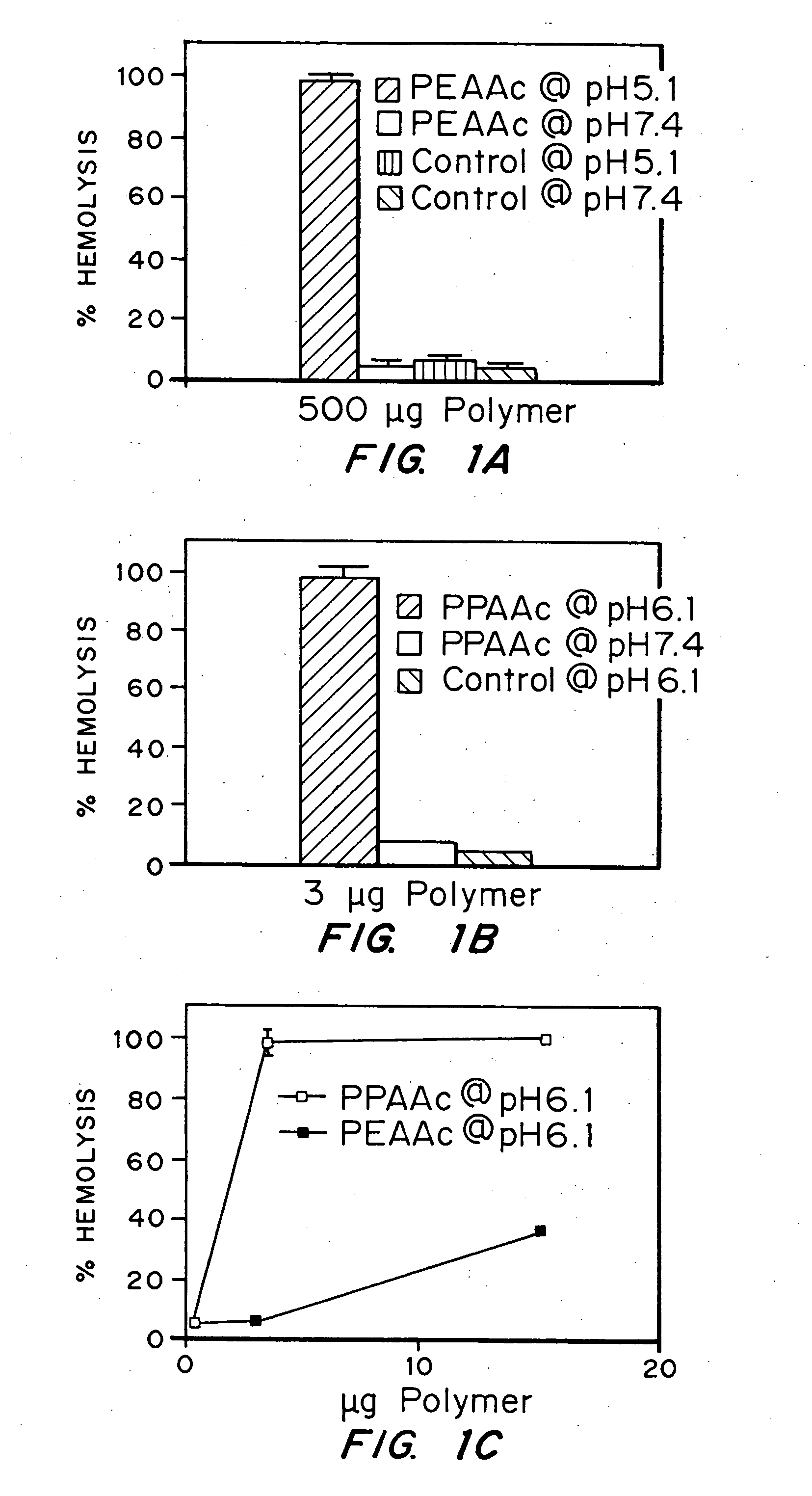
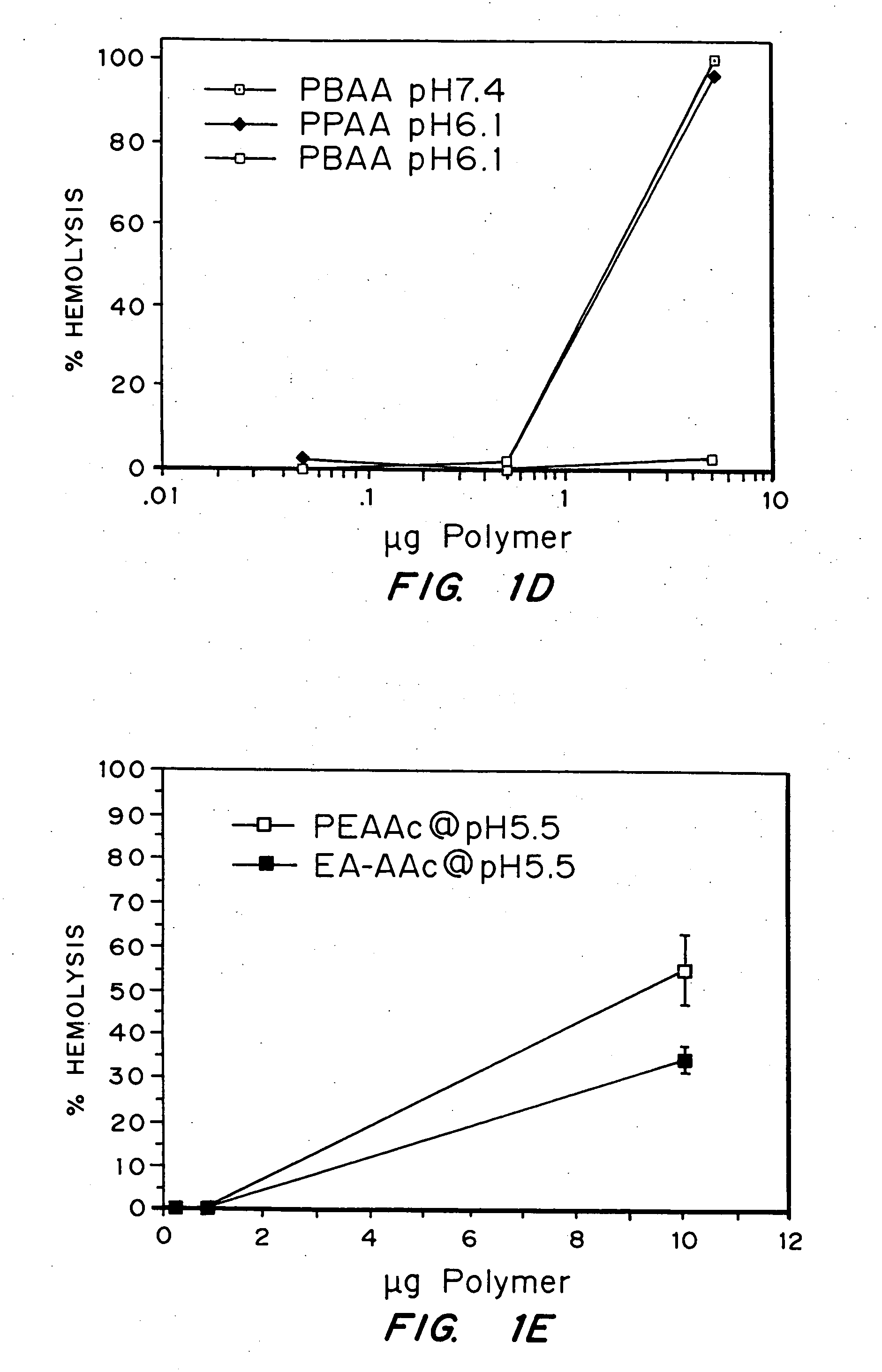
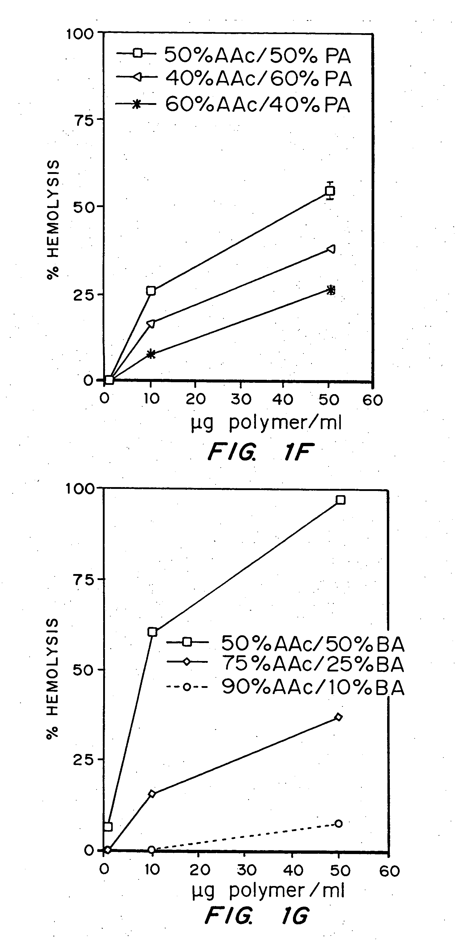
example 1
Evaluation of the pH sensitivity of PEAA-62K; PPAA.
[0089] Objective:
[0090] The objectives of this work were to determine if random copolymers of acrylic acid-butyl acrylate and acrylic acid-propyl acrylate have potential to act as endosomal releasing agents. This can be determined by measuring the hemolytic activity of the above polymers at endosomal pH (5.5) and physiologic pH (7.4).
[0091] Protocol:
[0092] (I) Polymer Synthesis: Polymers and random copolymers of acrylic acid-propyl acrylate and acrylic acid-butyl acrylate were synthesized by free radical polymerization at various monomer feed ratios, in bulk, using AIBN as the initiator. The comonomer feed ratios are indicated in the relevant figures. The polymers were purified by ether precipitation.
[0093] (II) Hemolysis Assay: Fresh human blood was isolated in EDTA containing vacutainers, washed three times with 150 mM NaCl, and resuspended at a concentration of 108 cells / ml in PBS buffer at either pH 5.5 or pH 7.4 or MES buf...
example 2
Comparison of the pH Sensitivity of EALA with that of an EALA / Polyacrylic Acid Conjugate
[0107] The ability of EALA to lyse erythrocytes was compared with that of an EALA / polyacrylic acid conjugate at a pH of 5.0 using approximately 107 red blood cells in 100 mM dibasic NaPO4, incubated at 37° C. for 20 minutes. A physical mixture of EALA and PAA was also tested.
[0108] The results are shown in FIG. 2. The EALA peptide by itself, as well as the physical mixture of EALA with PAA, demonstrated a negligible amount of lysis whereas the conjugate yielded about 70% lysis at a concentration of about 100 μg.
example 7
Comparison of the pH Sensitivity of PEAA with that of an IgG / PEAA Conjugate and that of IgG Alone
[0109] The ability of PEAA, an IgG / PEAA conjugate, and IgG alone to lyse erythrocytes was compared by performing a hemolysis assay at a pH of 5.0 using approximately 108 red blood cells in 100 mM dibasic sodium phosphate, and incubating at 37° C. for an hour. 1-ethyl-3-(3-dimethylaminopropyl)carbodiimide hydrochloride (EDC) was used to conjugate PEAA to rabbit immunoglobulin G (IgG). EDC reacts with the carboxyl groups to PEAA to form an amine-reactive intermediate, which then reacts with the lysine amine groups on IgG. The IgG was oxidized using 100 mM sodium periodate to yield reactive aldehyde groups on the carbohydrate moiety. Conjugation was realized via Schiff base formation between the end amine group of the amine-terminated PEAA and the aldehyde group on the IgG. This bond is reduced using 5 M sodium cyanoborohydride to yield a covalently bound conjugate of PEAA and IgG. The mol...
PUM
| Property | Measurement | Unit |
|---|---|---|
| pH | aaaaa | aaaaa |
| pH | aaaaa | aaaaa |
| pH | aaaaa | aaaaa |
Abstract
Description
Claims
Application Information