Systems and methods for analyzing pathologies utilizing quantitative imaging

a quantitative imaging and pathology technology, applied in image analysis, image enhancement, instruments, etc., can solve the problems of 45% misclassification, surgery does not benefit patients, and 45% of patients are not able to perform surgery, so as to improve image analysis, reduce signal noise, and improve image accuracy

Active Publication Date: 2018-11-15
ELUCID BIOIMAGING INC
View PDF1 Cites 49 Cited by
  • Summary
  • Abstract
  • Description
  • Claims
  • Application Information

AI Technical Summary

Benefits of technology

[0020]The present disclosure provides for improved image analysis. In particular, the systems and methods described herein relate to deblurring and segmentation of image data. The systems and methods advantageously account for and incorporate segmentation of biological analytes into a deblurring process for an image. Thus, the deblurring of the image may advantageously be optimized for enabling identification and quantitative analysis of one or more biological analytes based on underlying biological models for those analytes. The techniques described herein provide for significant improvements in the image deblurring and segmentation process which reduces signal noise and improves the accuracy of the image. Relative to existing techniques the present system and methods advantageously utilize unique optimization and tissue characteristics image models which may advantageously be informed by the underlying biology being analyzed, (for example by a biological model for the analytes). This provides for targeted deblurring and segmentation which is optimized for the applied image analytics.

Problems solved by technology

50% vascular surgeries do not benefit the patient, while some that need surgeries don't get them.
Results in 45% misclassification and inability to measure response to drugs or provide early intervention
Early intervention reduces mortality, but uncertain diagnosis is a constraint.
Lung cancer screening's potential for mortality reduction remains constrained by high burden of unnecessary costs.
Results in 25% false positive rate which limits benefit by burdening the system with errors and inefficiencies
Limitations of current image analysis tools lead to high rate of misclassification due to limitations in subjective and qualitative assessment using only a fraction of the information content in the images that are taken.
When, as is currently the case for cardiovascular diseases, the available markers for stratifying patients into risk categories are gross and unreliable, clinical trials of new therapeutic entities will inevitably be larger, more expensive and more likely to fail than would be the case with validated quantitative biomarkers for risk.
This is because poor assessment of risk leads to clinical trial populations that are insufficiently homogeneous and which include meaningful numbers of subjects not truly at elevated risk.
As a result, very large enrollments are necessary to achieve a statistically significant difference in outcomes between treatment and control groups even for a highly effective therapeutic.
In preclinical work, researchers need to analyze animal models to evaluate the efficacy of drugs on plaque severity before moving forward with expensive human clinical trials.
The principal limitations of current techniques include the difficulty in interpreting raw pixel reconstructed intensity values using simplistic thresholding operators.

Method used


Image

  • Systems and methods for analyzing pathologies utilizing quantitative imaging
  • Systems and methods for analyzing pathologies utilizing quantitative imaging
  • Systems and methods for analyzing pathologies utilizing quantitative imaging

Examples


Embodiment Construction

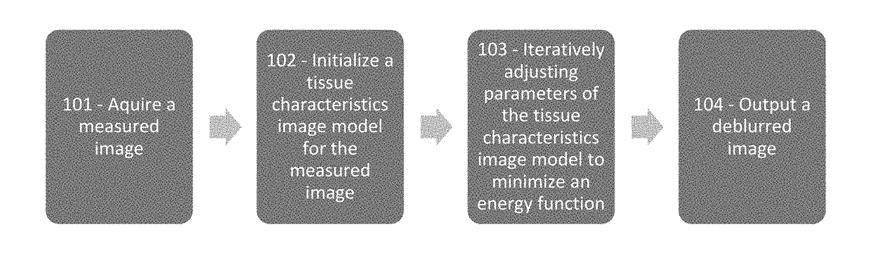
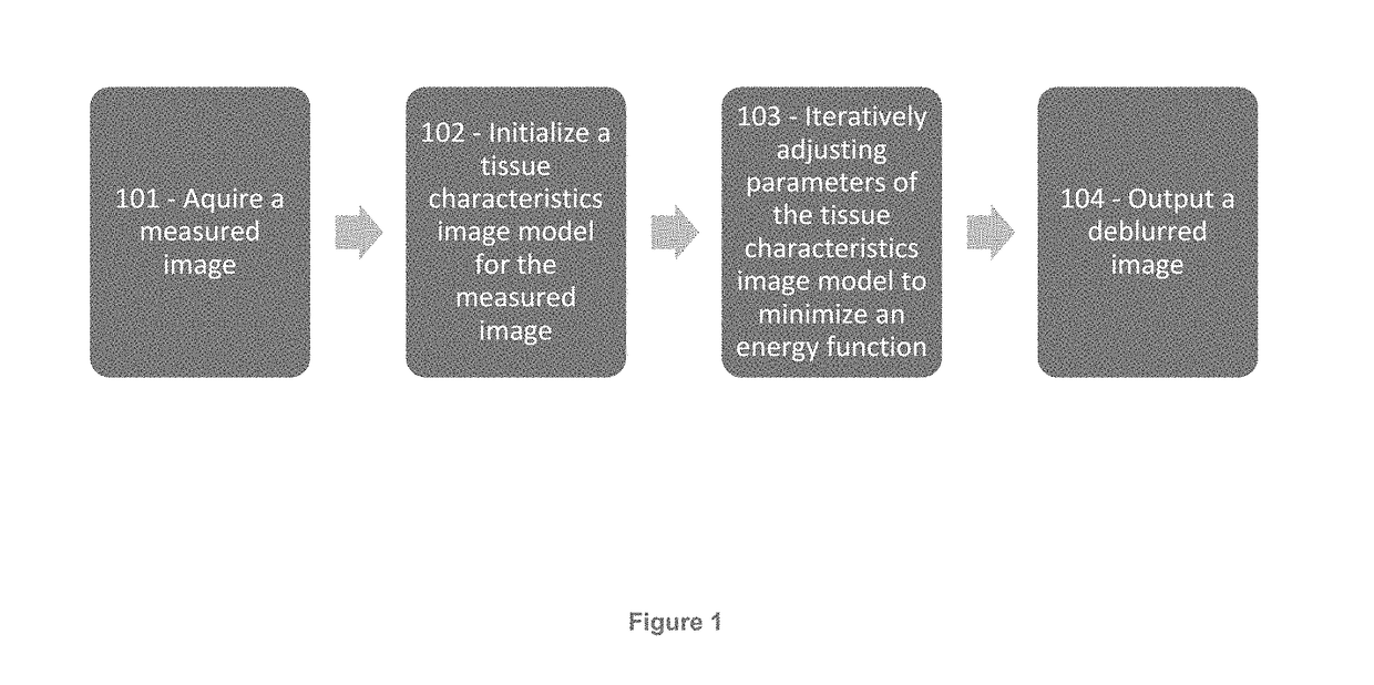
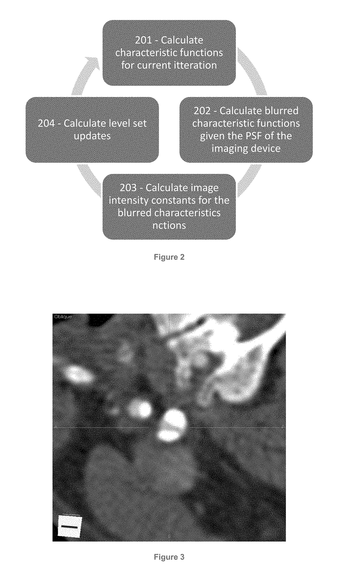
[0032]The present disclosure provides improved image interpretation capabilities which enable robust interpretation of medical images taking into account the non-idealities of the scanning process as well as a detailed knowledge of biological properties in combination. Example embodiments may relate to vascular analyte analysis of CT and / or MR images. Significant challenges to this analysis are 1) the analyte regions are extremely small, sometimes sub-voxel, 2) the scanner may have significant spatial blurring relative to the size of the analyte regions, and 3) the analyte regions are located very close to other high contrast edges (i.e., lumen-wall interface) whose blur (proportional to the contrast difference) may significantly change the intensity values. These conditions lead to blurring artifacts that are of an equal or greater image intensity magnitude than the analytes contrast with the surrounding tissue. These and other issues are addressed by way of the present disclosure....


PUM


Abstract

The present disclosure provides for improved image analysis via novel deblurring and segmentation techniques of image data. These techniques advantageously account for and incorporate segmentation of biological analytes into a deblurring process for an image. Thus, the deblurring of the image may advantageously be optimized for enabling identification and quantitative analysis of one or more biological analytes based on underlying biological models for those analytes. The techniques described herein provide for significant improvements in the image deblurring and segmentation process which reduces signal noise and improves the accuracy of the image. In particular, the system and methods described herein advantageously utilize unique optimization and tissue characteristics image models which are informed by the underlying biology being analyzed, (for example by a biological model for the analytes). This provides for targeted deblurring and segmentation which is optimized for the applied image analytics.

Description

CROSS REFERENCE TO RELATED APPLICATIONS[0001]The subject Application claims priority to U.S. Provisional Application Ser. No. 62 / 447,457, filed Jan. 18, 2017 and entitled “SYSTEMS AND METHODS FOR ANALYZING PATHOLOGIES UTILIZING QUANTITATIVE IMAGING.”STATEMENT REGARDING FEDERALLY SPONSORED RESEARCH OR DEVELOPMENT[0002]This work supported in part by ID 1353532 awarded by NSF and the government may have certain rights to the work ID 10025457 awarded by NIH.BACKGROUND OF THE INVENTION[0003]The present disclosure related to quantitative imaging and analytics. More specifically, the present disclosure relates to systems and methods for analyzing pathologies utilizing quantitative imaging.[0004]Imaging, particularly with safe and non-invasive methods, represents the most powerful methods for locating the disease origin, capturing its detailed pathology, directing therapy, and monitoring progression to health. Imaging is also an extremely valuable and low cost method to mitigate these human...

Claims


Application Information

Patent Timeline
Patent Type & Authority Applications(United States)
IPC IPC(8): G06T5/00G06T7/136G06T7/11G06T7/194G06T7/00G06K9/46G06V10/26
CPCG06T2207/10081G06T2207/10088G06K9/4647G06T5/003G06T7/136G06T2207/20076G06T7/194G06T7/0012G06K2209/05G06T2207/30101G06T2207/20152G06T7/11G06T2207/10072G06T2207/20161G06T7/143G06T2207/10132G06T2207/20036G06T2207/20081G06T2207/30024G06T2207/30096G06V10/26G06V10/7557G06V2201/03
Inventor PAIK, DAVID S.BUCKLER, ANDREW J.JOHNSON, KJELLMA, XIAONANMOULTON, KEITH A.
Owner ELUCID BIOIMAGING INC
Why Eureka
Try Eureka