
Tissue Processor

a tissue processor and tissue technology, applied in the field of tissue processors, can solve the problems of not being practicable, not being able to achieve the real workflow, and forcing for a compromise between speed and quality of process, so as to avoid contamination of operators by containers, improve processing efficiency, and reduce processing costs

Active Publication Date: 2019-01-31
MILESTONE SRL
View PDF5 Cites 1 Cited by
  • Summary
  • Abstract
  • Description
  • Claims
  • Application Information

AI Technical Summary

Benefits of technology

The invention is a tissue processor that can automatically process single tissue carriers without being dependent on other carriers being processed simultaneously. The processor generates an independent flow for each carrier, resulting in more efficient processing and higher quality. The processor also allows for individualized processing, reducing the need for buffer containers and minimizing fume exposure. The processor includes a control unit that can control the heater, stirrer, and / or pump for performing histological processes in each container. The stirrer helps to prevent temperature stratification and enhance the reaction of the liquid with the tissue carrier. Overall, the invention provides a better and more effective way to process tissue carriers.

Problems solved by technology

These processes must accommodate the needs of different tissue specimens, forcing for a compromise between speed and quality of the process.
To avoid compromises, grouping similar tissue types (e.g., same organ type and same thickness) in the same batch is required, but it is not usually feasible in the real workflow: the collection of tissue specimens from patients is not scheduled and sorted based on tissue type / size basis.
However, it is not practically feasible: the number of tissues / tissue specimens processed in two or three hours with a continuous flow tissue processor used with “single-tissue batches” would be about 10 or 15 or less, instead of several hundreds of tissue specimens.
For this reason, this way would be economically unprofitable and is therefore disadvantageous.
Because of the FIFO queuing, there is no possibility to manage a priority for urgent tissue specimens: if a process is already in execution, the urgent tissue carrier or tissue specimen must wait to be processed within the next batch.
Moreover, a previous ongoing batch process that requires a long time could further slowdown the urgent tissue specimens, since the urgent tissue specimen process cannot go over the ongoing processes.
This method introduces useless delays, workflow pikes and the need of a proper buffer space to collect the prepared tissue specimens.

Method used


Image

  • Tissue Processor
  • Tissue Processor
  • Tissue Processor

Examples


Embodiment Construction

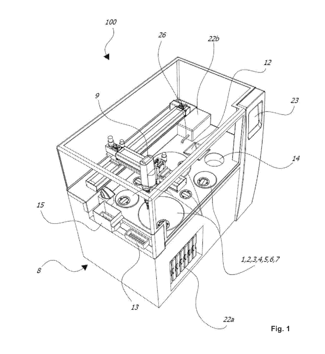
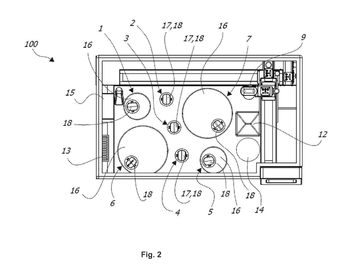
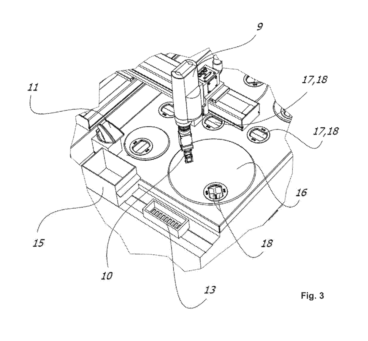
[0038]In the following, the invention is described exemplarily with reference to the enclosed figures, in which

[0039]FIG. 1 is a perspective view of an exemplary embodiment of the tissue processor according to the invention;

[0040]FIG. 2 is a plane view of the tissue processor shown in FIG. 1;

[0041]FIG. 3 is a top view of the tissue processor shown in FIG. 1;

[0042]FIG. 4 is a functional diagram of a part of the tissue processor shown in FIG. 1;

[0043]FIG. 5 is an exploded view of a preferred tissue carrier;

[0044]FIG. 6 is a side view of the tissue carrier shown in FIG. 5;

[0045]FIG. 7 is a table showing an exemplary protocol for processing different tissue specimens by the tissue processor;

[0046]FIG. 8 is a table showing an exemplary timeline for concurrently processing the different tissue specimens of FIG. 7.

[0047]FIGS. 1 to 3 show an exemplarily tissue processor 100 for automatically processing (preferably at least processing including embedding) histological tissue specimens. The t...


PUM


Abstract

The invention relates to a tissue processor (100) for automatically processing histological tissue specimens, the tissue processor (100) comprising a plurality of containers (1, 2, 3, 4, 5, 6, 7) each being provided for a respective histological process for processing at least one tissue carrier, a robotic arm (9) for handling a tissue carrier between the containers (1, 2, 3, 4, 5, 6, 7) for being processed in each of the containers (1, 2, 3, 4, 5, 6, 7), and a control unit for controlling the histological process in each of the containers (1, 2, 3, 4, 5, 6, 7) and for controlling the robotic arm (9), wherein the control unit is configured such that the robotic arm (9) handles a tissue carrier between the containers (1, 2, 3, 4, 5, 6, 7) while the histological process in each of the containers (1, 2, 3, 4, 5, 6, 7) continues in an uninterrupted fashion, so that the execution of the process for each tissue carrier is independent from the loading order and / or process duration of other tissue carriers being processed in the containers (1, 2, 3, 4, 5, 6, 7).

Description

1. FIELD OF THE INVENTION[0001]The present invention relates to a tissue processor for automatically processing histological tissue specimens and a method using such a tissue processor for automatically processing histological tissue specimens.2. TECHNICAL BACKGROUND[0002]Usually, tissue specimens or tissue samples are taken at a hospital and subsequently transferred to a laboratory or the like for histological examination. For a histological examination of human and / or animal tissue specimens, the tissue specimens are collected from the patients by a clinician in an ambulatory environment (biopsies) or by surgeons during a surgery. After that, the tissue specimens are placed in suitable transportation carriers such as a container. The transportation carrier is typically filled with a fixative such as formalin and sent to the histology laboratory for final examination.[0003]After the histology laboratory has received the tissue specimens, the first operation performed by a histotech...

Claims


Application Information

Patent Timeline
IPC IPC(8): G01N1/30G01N35/00B01L3/00G01N1/36
CPCG01N1/30G01N35/0099G01N35/0095B01L3/523G01N1/36G01N2035/00346B01L2300/04B01L2300/045G01N1/31G01N35/00732G01N35/0092G01N2035/00851
Inventor MINUTI, MATTEOVISINONI, FRANCESCO
Owner MILESTONE SRL
Learn More