Method and system for assessing a haemodynamic parameter

a haemodynamic parameter and parameter technology, applied in the field of assessing haemodynamic parameters, can solve the problems of inability to reconstruct or characterize neighboring vessels, and inability to accurately assess the physiological significance of lesions in coronary angiography. achieve the effect of accurate assessment of haemodynamic parameters, accurate confidence value of assessment, and high confidence valu

Active Publication Date: 2019-05-23
SIEMENS HEALTHCARE GMBH
View PDF0 Cites 7 Cited by
  • Summary
  • Abstract
  • Description
  • Claims
  • Application Information

AI Technical Summary

Benefits of technology

[0039]Multiple image quality values may be determined for different regions or parts of any single angiographic image or view. If, for example, only one vessel segment centrally located in an angiographic image is in focus (e.g., is imaged sharply), a relatively high confidence value may be assigned to a geometric feature derived or extracted from that particular, central vessel segment. At the same time, another geometric feature may be extracted from a different part of the same angiographic image, for example, for a neighboring healthy vessel segment that is out of focus. Since oftentimes in angiographic imaging no particular attention is paid to an image quality for vessels or segments other than those containing or immediately surrounding a stenosis or lesion of interest, determining and using an image quality metric with sub-image level accuracy may be particularly advantageous for the presently described method. This may be the case because it may ultimately result in a more accurate assessment of the haemodynamic parameter and / or a more accurate confidence value of the assessment.
[0040]In a further development, based on the provided angiographic images, it is automatically determined or computed what additional input data could or would improve the calculated confidence value. A corresponding suggestion is then issued. If, for example, a specific geometric feature has a confidence value below a predetermined threshold, then it may be suggested to acquire an additional angiographic image of a specific part of the region of interest corresponding to that geometric feature. This may limit the number of additional images that are to be acquired to achieve an assessment of the haemodynamic parameter with at least a minimum confidence value. Therefore, any additional strain or exposure for the patient may be minimized. The suggestion may also contain or provide specific parameters for a suggested image acquisition, such as a specific or minimal dosage of a contrast agent and / or a specific angulation and / or an imaging modality to be used. The suggestion may be issued automatically or only if a confidence value of the assessment of the haemodynamic parameter, or of one or more geometric features is below a predefined threshold. It may be possible for medical personnel to request a suggestion at any time, regardless.

Problems solved by technology

Nonetheless, coronary angiography has some intrinsic limitations and shortcomings.
One of the most significant limitations of coronary angiography is an inability to accurately assess the physiological significance of lesions.
In many cases, this limits the possibility of reconstructing or characterizing neighboring vessels to obtain a complete representation or reconstruction or model of the vessel tree surrounding an imaged feature of interest, such as a stenosis.
In these cases or conditions, it is not possible to fully represent or reconstruct the complete 3D geometry and topology of the vessel tree or arterial tree.
The required additional data is, however, not reliably available for every patient.
Therefore, to fully represent or reconstruct the vessel tree in a region of interest, additional images would have to be acquired at the cost of additional exposure of the patient to radiation and contrast medium as well as additional expenditure of time and money.
Even if additional images are available or acquired, it may be impossible or extremely difficult to combine all data into a single complete and correct 3D representation.
One difficulty lies in the needed registration between different datasets, which, for example, may have been acquired at different points in time and / or using different imaging equipment.
In this case, combining the respective data or models into a single cohesive and consistent 3D model may, at least for practical purposes, be impossible without proper position tracking at all times.

Method used


Image

  • Method and system for assessing a haemodynamic parameter
  • Method and system for assessing a haemodynamic parameter
  • Method and system for assessing a haemodynamic parameter

Examples


Embodiment Construction

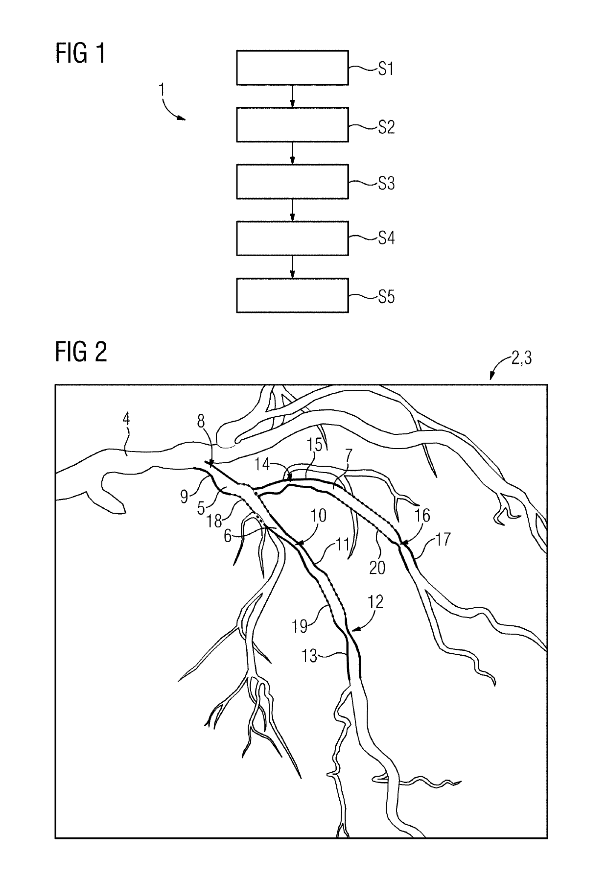
[0052]FIG. 1 schematically shows a flow chart 1 of one embodiment of a method for assessing a haemodynamic parameter such as a fractional flow reserve (FFR) based on multiple angiographic images. In a process act S1, the angiographic images of a vascular region of interest are acquired. This may include actually taking the images using a medical imaging system or device and / or may include accessing a data storage device on which the angiographic images are stored. The region of interest may contain or include multiple different sub-regions, parts, or portions. The angiographic images may contain or enable one or more complete, partial, or incomplete views of each of these parts or portions.

[0053]In a process act S2, a 3D representation or reconstruction of at least a first portion of the region of interest is obtained based on the angiographic images. Geometric features are extracted from complete or partial views of a respective anatomical structure of interest (e.g., from the 3D r...


PUM


Abstract

A method and a corresponding system for assessing a haemodynamic parameter for a vascular region of interest of a patient based on angiographic images are provided. After acquiring multiple angiographic images, a three dimensional (3D) representation of at least a first portion of the respective region of interest is performed, and geometric features are extracted from complete or partial views. Additional geometric features are extracted from partial incomplete views. A complete set of 3D geometric features for an anatomical structure, such as a vessel tree, is then generated by combining the extracted geometric features and estimating any missing geometric features. Using the complete set of 3D geometric features, a feature-based assessment of the haemodynamic parameter, such as a fractional flow reserve, is then performed.

Description

[0001]This application claims the benefit of EP17464012, filed on Nov. 22, 2017, which is hereby incorporated by reference in its entirety.BACKGROUND[0002]The present embodiments relate to assessing a haemodynamic parameter.[0003]Today, angiography is a well-established medical imaging technique. Coronary angiography, for example, is considered the gold standard for anatomical assessment of coronary artery disease. Nonetheless, coronary angiography has some intrinsic limitations and shortcomings. One of the most significant limitations of coronary angiography is an inability to accurately assess the physiological significance of lesions. Subject-specific physiology modeling may address this limitation. The approach includes determining the geometry features of the patient-specific arterial tree, and subsequent use of a computational model designed to predict functional diagnostic indices, such as FFR, as a function of the geometry.[0004]US 2014 / 0058715A1 discloses a method and a sys...

Claims


Application Information

Patent Timeline
Patent Type & Authority Applications(United States)
IPC IPC(8): A61B6/00G16H30/20G06N20/00G06N3/08
CPCA61B6/5217A61B6/5229A61B6/504A61B6/507A61B6/481G16H30/20G06N20/00G06N3/08G06T7/0012G06T2207/10021G06T2207/10116G06T2207/30104
Inventor PASSERINI, TIZIANOITU, LUCIAN MIHAIREDEL, THOMASSHARMA, PUNEET
Owner SIEMENS HEALTHCARE GMBH
Why Eureka
Try Eureka