
System and Method for Monitoring Airflow in a Subject's Airway with Ultrasound

a technology of airflow monitoring and subject airway, applied in the field of system and method for monitoring airflow in a subject's airway with ultrasound, can solve the problems of respiratory failure, impede the timely implementation of rescue measures, and high morbidity and mortality rates of spontaneous breathing patients

Pending Publication Date: 2019-06-06
WISCONSIN ALUMNI RES FOUND
View PDF2 Cites 6 Cited by
  • Summary
  • Abstract
  • Description
  • Claims
  • Application Information

AI Technical Summary

Benefits of technology

The present invention provides systems and methods for non-invasively monitoring airflow in a subject's airway using ultrasound. By acquiring Doppler ultrasound signals from anatomical regions in the airway, the technology can compare them to baseline data to identify when there is a significant deviation that could impact breathing. An alarm is then triggered to alert the user. This offers a safe and non-invasive way to monitor airflow and can be used in a variety of medical settings.

Problems solved by technology

Despite technological advancements and improved anesthetic agents leading to an increased safety profile of anesthesia and sedation, morbidity and mortality rates remain high in spontaneously breathing patients.
One of the primary reasons morbidity and mortality rates remain high is the delayed detection of early respiratory compromise and / or failure, which impedes the timely implementation of rescue measures.
Respiratory failure can occur within minutes to seconds, or it can develop gradually, both of which are difficult to predict.
The end results of respiratory compromise and / or failure is insufficient oxygen to the brain and heart, leading to grave consequences including permanent neurological and cardiac damage, or even death.
Currently available monitoring devices and techniques continue to be ineffective at alerting to subtle airflow changes in the early stages of respiratory compromise and / or failure.
Patients under sedation can experience decreased respiratory effort, decreased respiratory rate, and varying degrees of tissue laxity, which can lead to airway obstruction and both of which are difficult to assess.
Any combination of these mechanisms can lead to diminished airflow and / or changes in airflow, which prevents gas exchange for oxygen and carbon dioxide.
Accurately monitoring for respiratory compromise and / or failure is especially difficult in pediatric patients, whose anatomy makes maintaining airway and respiratory homeostasis challenging during inhalation induction for anesthesia or sedation.
For example, pediatric patients experience higher rates of tongue obstruction during sedation and anesthesia due to their larger tongue size in relation to their oral cavity.
Unrecognized and delayed respiratory support requiring skilled airway maneuvers (e.g., hand-bag-ventilation) will lead to severe desaturation followed by bradycardia, which can be fatal for the patient.
As mentioned above, the currently available methods for monitoring respiratory compromise and / or failure are ineffective at directly measuring airflow changes in non-intubated patients and spontaneously breathing patients, thereby rendering the methods unreliable for accurately and timely detecting early respiratory compromise and / or failure.
However, measuring end-tidal CO2 has inherent limitations because it is an indirect measurement of alterations in airflow and becomes increasingly inaccurate in non-intubated patients due to the lack of a closed circuit.
As a consequence, the measured data are difficult for the practitioner to interpret in spontaneously breathing patients, which often leads to delays in the treatment of early airway compromise and / or respiratory failure.
However, pulse oximetry does not directly monitor respiration and hence does not monitor ventilation.
For example, an obstructed airway will decrease oxygen flow and hence oxygen supply to the body, leading to desaturation (i.e., a drop in oxygen).
The limitation of pulse oximetry is a delayed response to desaturation, resulting in a time lag in the detection of hypoxic events, particularly in the presence of supplemental oxygen.
Moreover, ECG-monitoring does not provide real-time measurements necessary to timely identify early airway compromise and / or failure.
This measurement technique, however, is very susceptible to erroneous readings secondary to motion artifact.
In the setting of a mass casualty (e.g., in battlefield medicine or natural or human disasters) triage is very challenging.

Method used


Image

  • System and Method for Monitoring Airflow in a Subject's Airway with Ultrasound
  • System and Method for Monitoring Airflow in a Subject's Airway with Ultrasound
  • System and Method for Monitoring Airflow in a Subject's Airway with Ultrasound

Examples


Embodiment Construction

[0023]Described here are systems and methods for monitoring airflow changes in a subject's airway. This monitoring may be performed before, during, or following a procedure, or may be used as a general patient monitoring tool. For example, the systems and methods of the present disclosure can be used to monitor signs of airway obstruction or respiratory compromise and / or failure during timeframes where patients have ongoing or residual sedation or anesthetics in their system. The systems and methods can also be implemented for quantitatively measuring airflow and related parameters. As one example, the systems and methods of the present disclosure can be used to quantitatively measure respiratory rate in real-time. In addition, the systems and methods described in the present disclosure provide an improved accuracy because they are capable of detecting actual breaths as opposed to attempted breaths, such as in upper airway obstruction when the chest wall continues to make efforts ag...


PUM


Abstract

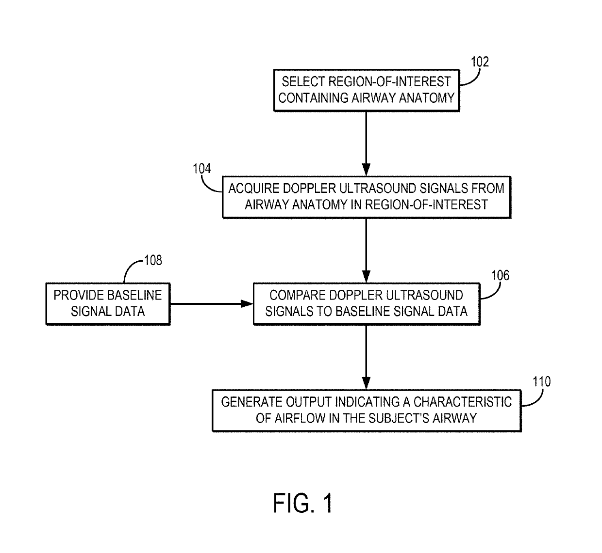
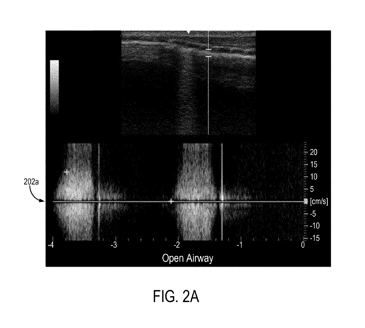
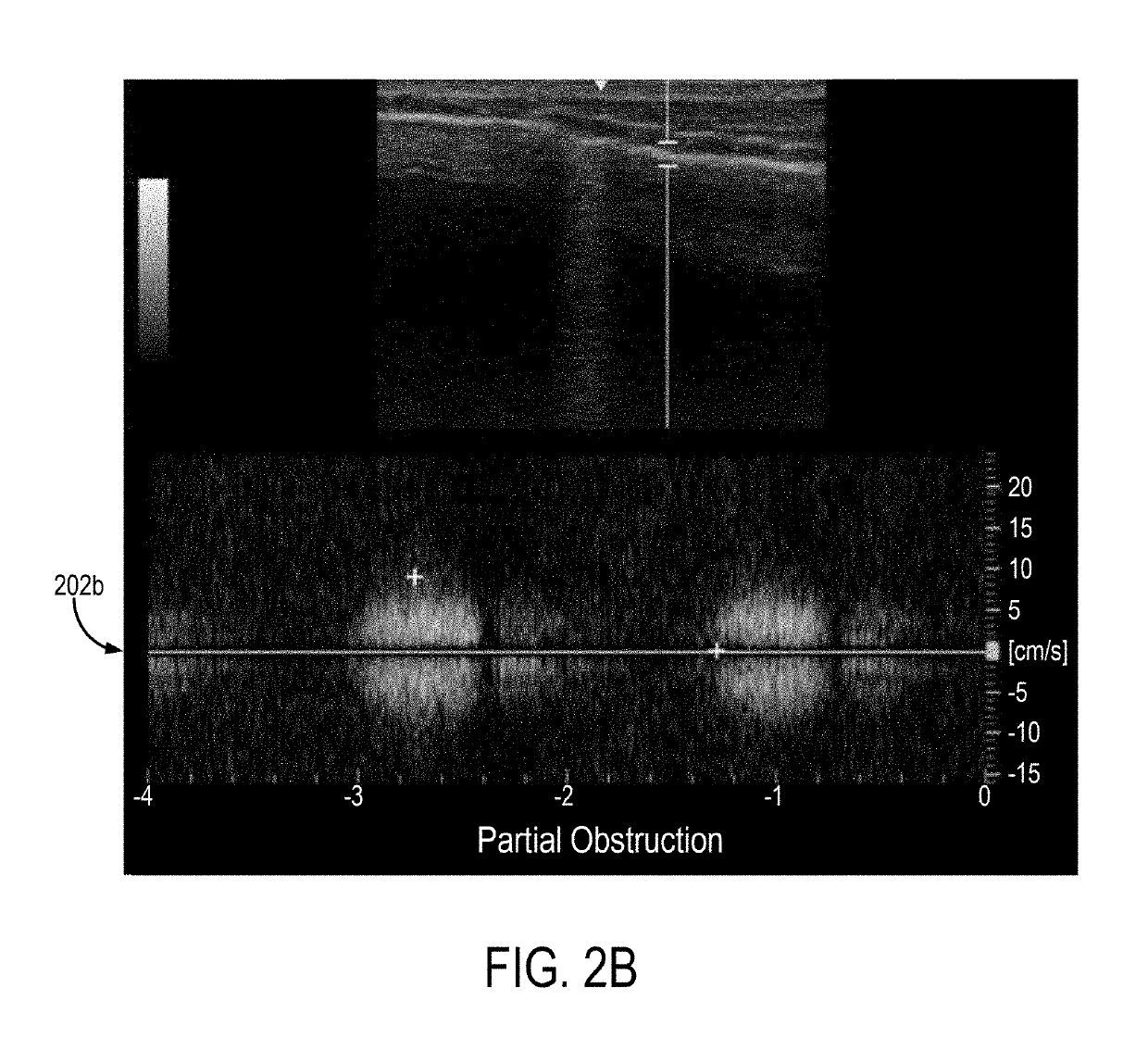
Described here are systems and methods for monitoring airflow changes in a patient's airway during a medical procedure or as a general patient monitoring tool. Doppler ultrasound signals are acquired from an anatomical region within the patient's airway (e.g., a tracheal wall, a cricothyroid ligament, other connective or cartilaginous tissue within the trachea, larynx, or pharynx) and parameters from those Doppler ultrasound signals are compared to baseline parameters, which may include inputting Doppler ultrasound signals to a suitably trained deep learning model or other machine learning algorithm. When a threshold change is detected, an alarm can be provided to a user to indicate respiratory compromise and / or failure, which can include early airway compromise, airway failure, and / or airway obstruction.

Description

CROSS-REFERENCE TO RELATED APPLICATIONS[0001]This application is a continuation-in-part of claims the benefit of International Application No. PCT / US2017 / 052233, filed on Sep. 19, 2017, which claims the benefit of U.S. Provisional Application No. 62 / 396,339, filed on Sep. 19, 2016, both of which are herein incorporated by reference in their entirety.BACKGROUND[0002]In the past few decades, advances in monitoring technology have contributed to a heightened sense of safety during medical procedures. Despite technological advancements and improved anesthetic agents leading to an increased safety profile of anesthesia and sedation, morbidity and mortality rates remain high in spontaneously breathing patients. One of the primary reasons morbidity and mortality rates remain high is the delayed detection of early respiratory compromise and / or failure, which impedes the timely implementation of rescue measures. Respiratory failure can occur within minutes to seconds, or it can develop gradu...

Claims


Application Information

Patent Timeline
IPC IPC(8): A61B8/08A61B8/00A61B5/087A61B5/00
CPCA61B8/08A61B8/488A61B8/469A61B5/087A61B5/746A61B5/7267A61B5/7405A61B5/742A61B8/5207A61B5/0816A61B5/085A61B5/4821A61B7/003
Inventor BILEN-ROSAS, GUELAYROSAS, HUMBERTO GERARDO
Owner WISCONSIN ALUMNI RES FOUND
Learn More