Method for predicting efficacy of immune checkpoint inhibitors in cancer patients

a technology cancer patients, which is applied in the field of predicting can solve the problems of inaccurate experimental models, and inability to accurately predict the efficacy of immune checkpoint inhibitors in cancer patients

Inactive Publication Date: 2019-06-06
BEIGENE
View PDF0 Cites 1 Cited by
  • Summary
  • Abstract
  • Description
  • Claims
  • Application Information

AI Technical Summary

Benefits of technology

[0014]Disclosed herein is a fast and ex vivo method for predicting clinical efficacy of an immune checkpoint inhibitor or a combination of two or more thereof or a combination of an immune checkpoint inhibitor and other antitumor drugs or a method for screening an immune checkpoint inhibitor or a combination of two or more thereof or a combination of an immune checkpoint inhibitor and other antitumor drugs effective in the treatment of a cancer of interest and the new use of an BiTE antibody for predicting clinical efficacy of an immune checkpoint inhibitor or for drug screening.

Problems solved by technology

Although many patients seem to benefit from the new treatments, results are not uniform for all patients; thus, there is a critical need for developing biomarkers that permit identification of patients that will benefit from a particular treatment.
The development of accurate experimental models of cancer is always a key challenge for both the investigation of basic cancer biology and the development of novel therapeutic modalities.
It is well recognized that one of the key challenges in developing immune-oncology agents is to evaluate their activities as single agent or in combinations by predictive and robust preclinical models.
On the other hand, traditional xenograft or patient derived xenograft (PDX) models do not have representative TME for human tumors as typically immune-deficient mice are required for xenograft models.
This requirement has limited utilities of syngeneic mouse models in investigating and comparing immune-oncology agents.
However, there are technical challenges in this approach that have limited the progresses in this field.
One of the main hurdles for an ex vivo or in vitro system is that the activation of the immune system via tumor associated antigens (TAAs) is either missing or insufficient to induce strong antitumor immune responses.
Therefore, it is very hard to detect changes of the anti-tumor immune response.

Method used


Image

  • Method for predicting efficacy of immune checkpoint inhibitors in cancer patients
  • Method for predicting efficacy of immune checkpoint inhibitors in cancer patients
  • Method for predicting efficacy of immune checkpoint inhibitors in cancer patients

Examples


example 1

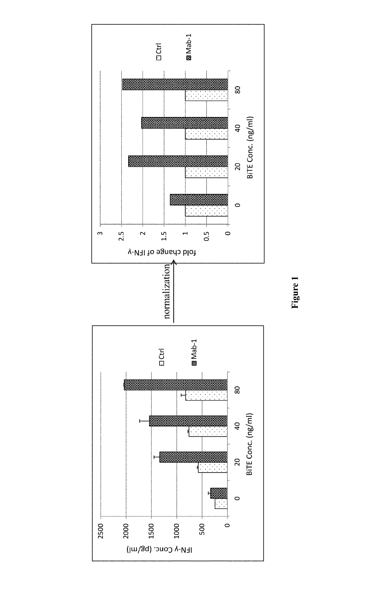
[0145]Primary tumor cells from a patient with adenocarcinoma of the esophagogastric junction cultured on feeder cells were co-cultured with pre-activated allogeneic human PBMCs, with different concentration of EpCAM×CD3 BiTE and with or without different anti-PD-1 antibody drugs (0.1 μg / mL), which are Mab-1 and cultured for 24 hours. Human IFN-γ in the supernatant of the co-culture system was measured and as the readout (See FIG. 1). The IFN-γ concentration of each BiTE concentration group was normalized to fold change by antibody treatment group concentration dividing by control group concentration.

[0146]At the group without BiTE, the fold changes of anti-PD-1 antibody treatment groups were all smaller than 1.3, which can be explained by the fact that PBMCs and tumor cells only physically contacted and there was no or litter immune synapse formation between PBMCs and tumor cells, PD-1 signal wasn't activated, little, if any. While at the groups with BiTE, the fold changes of anti-P...

example 2

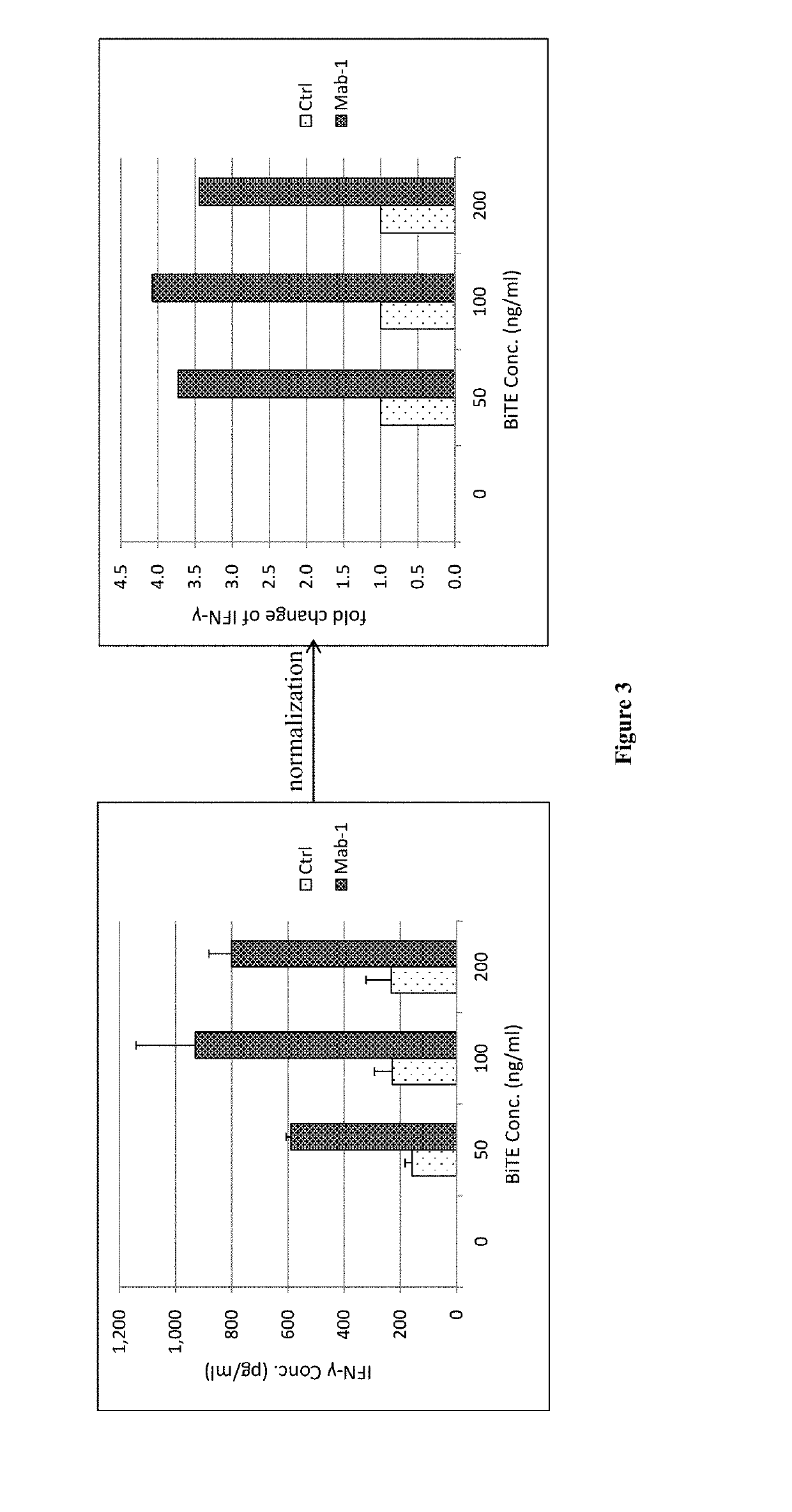
[0148]Primary tumor cells from a patient with colorectal cancer cultured in Matrigel® were co-cultured with non pre-activated allogeneic human PBMCs, with different concentration of EpCAM×CD3 BiTE and with or without 1 μg / mL of Mab-1 and cultured for 72 hours. Human IFN-γ in the supernatant of the co-culture system was measured and as the readout (See FIG. 3). The IFN-γ concentration of each BiTE concentration group was normalized to fold change by antibody treatment group concentration dividing by control group concentration.

[0149]At the groups without BiTE, Human IFN-γ can't be detected by ELISA. While at the groups with BiTE, the fold changes of Mab-1 treatment groups were all bigger than 1.3 and almost 3.5 times of control groups, which indicated that the primary tumor cells responded to Mab-1 treatment and predicted that there is a substantial possibility that this cancer patient will respond to the tested anti-PD-1 antibody. So, the primary tumor cells cultured in Matrigel® re...

example 3

[0150]Primary tumor cells from a patient with colon cancer cultured in Matrigel® were co-cultured with non pre-activated allogeneic human PBMCs, with different concentration of EpCAM×CD3 BiTE and with or without an anti-PD-1 antibody named Mab-1 (1 μg / mL), and the combination of Mab-1 (1 μg / mL) and 5-(((1R,1aS,6bR)-1-(6-(trifluoromethyl)-1H-benzo[d]imidazol-2-yl)-1a,6b-dihydro-H-cyclopropa[b]benzofuran-5-yl)oxy)-3,4-dihydro-1,8-naphthyrdin-2(1H)-one Sesqui-Maleate (Compound 1, 300 nM) (named as Mab-1+Compound 1, for short), and cultured for 72 hours. Human IFN-γ in the supernatant of the co-culture system was measured and as the readout (See FIG. 4). The IFN-γ concentration of each BiTE concentration group was normalized to fold change by antibody treatment group concentration dividing by control group concentration. Primary tumor cells cultured in Matrigel® responded to anti-PD-1 antibody.

[0151]At the groups without BiTE, Human IFN-γ can't be detected by ELISA. While at the groups ...


PUM

PropertyMeasurementUnit
molecular weightsaaaaaaaaaa
concentrationaaaaaaaaaa

Abstract

Disclosed is a method for predicting clinical response of a cancer patient to treatment with an immune checkpoint inhibitor, or a combination of two or more thereof, or with a combination of an immune checkpoint inhibitor and other antitumor drugs, by introducing bispecific T cell engager (BiTE) into the platform of lymphocytes containing T cells in vitro, which are cultured on feeder cells or in Matrigel®.

Description

CROSS REFERENCE TO RELATED APPLICATIONS[0001]This application is a U.S. National Stage Application under 35 U.S.C. § 371 of International Application No. PCT / CN2017 / 095144, filed on Jul. 31, 2017, which claims the benefit of priority to International Application No. PCT / CN2016 / 093917 filed on Aug. 8, 2016, the entire contents of each of which are hereby incorporated by reference.FIELD OF THE INVENTION[0002]Disclosed herein is a method for predicting clinical efficacy of an immune checkpoint inhibitor or a combination of two or more thereof or a combination of an immune checkpoint inhibitor and other antitumor drugs, or a method for screening an immune checkpoint inhibitor or a combination of two or more thereof or a combination of an immune checkpoint inhibitor and other antitumor drugs effective in the treatment of a cancer of interest and the new use of an BiTE antibody for predicting clinical efficacy of an immune checkpoint inhibitor or for drug screening. The methods disclosed ...

Claims


Application Information

Patent Timeline
Patent Type & Authority Applications(United States)
IPC IPC(8): G01N33/574C12Q1/02C07K16/28
CPCG01N33/574C12Q1/025C07K16/2818G01N2500/02G01N2500/10A61P35/00C07K2317/76
Inventor LUO, LUSONGDAI, CHENGLIANGSUN, ZHIJIAN
Owner BEIGENE
Why Eureka
Try Eureka