Methods and devices for the treatment of pulmonary disorders

a technology for pulmonary disorders and pulmonary artery disease, applied in the field of can solve the problems of existing lung artery reduction devices, devices that do not represent optimal solutions for lung artery reduction, and devices that have yet to enter widespread use, so as to reduce the risk of device implantation, increase the potential speed of device placement, and reduce the effect of arm bias

Inactive Publication Date: 2019-10-24
EOLO MEDICAL INC
View PDF0 Cites 0 Cited by
  • Summary
  • Abstract
  • Description
  • Claims
  • Application Information

AI Technical Summary

Benefits of technology

[0016]The inventors have conceived of and disclose herein, implantable lung volume reducing devices and medical techniques for implanting lung volume reduction devices through the trachea and bronchi, using minimally-invasive deployment and surgical techniques. The lung reducing devices may be used to reduce the volume of one or more lung, thereby increasing the elastic recoil of the remaining lung volume.
[0035]In accordance with these and the further aspects of the present invention, a method and device is described for reducing the volume of a lung. The present invention provides advantages of a minimally invasive procedure for alleviating at least some of the symptoms associated with COPD and emphysema without the risk and complications associated with conventional LVRS surgery.

Problems solved by technology

The devices have yet to enter into widespread use.
While the lack of use is at least partially due to the lack of government approval in the United States, it is posited that existing lung reducing devices and the techniques to implant the device do not represent optimal solutions for lung reduction.
These devices may also delay closure of the small airways in the lung during a breath and lower the Residual Volume (RV) in the lung.

Method used


Image

  • Methods and devices for the treatment of pulmonary disorders
  • Methods and devices for the treatment of pulmonary disorders
  • Methods and devices for the treatment of pulmonary disorders

Examples


Embodiment Construction

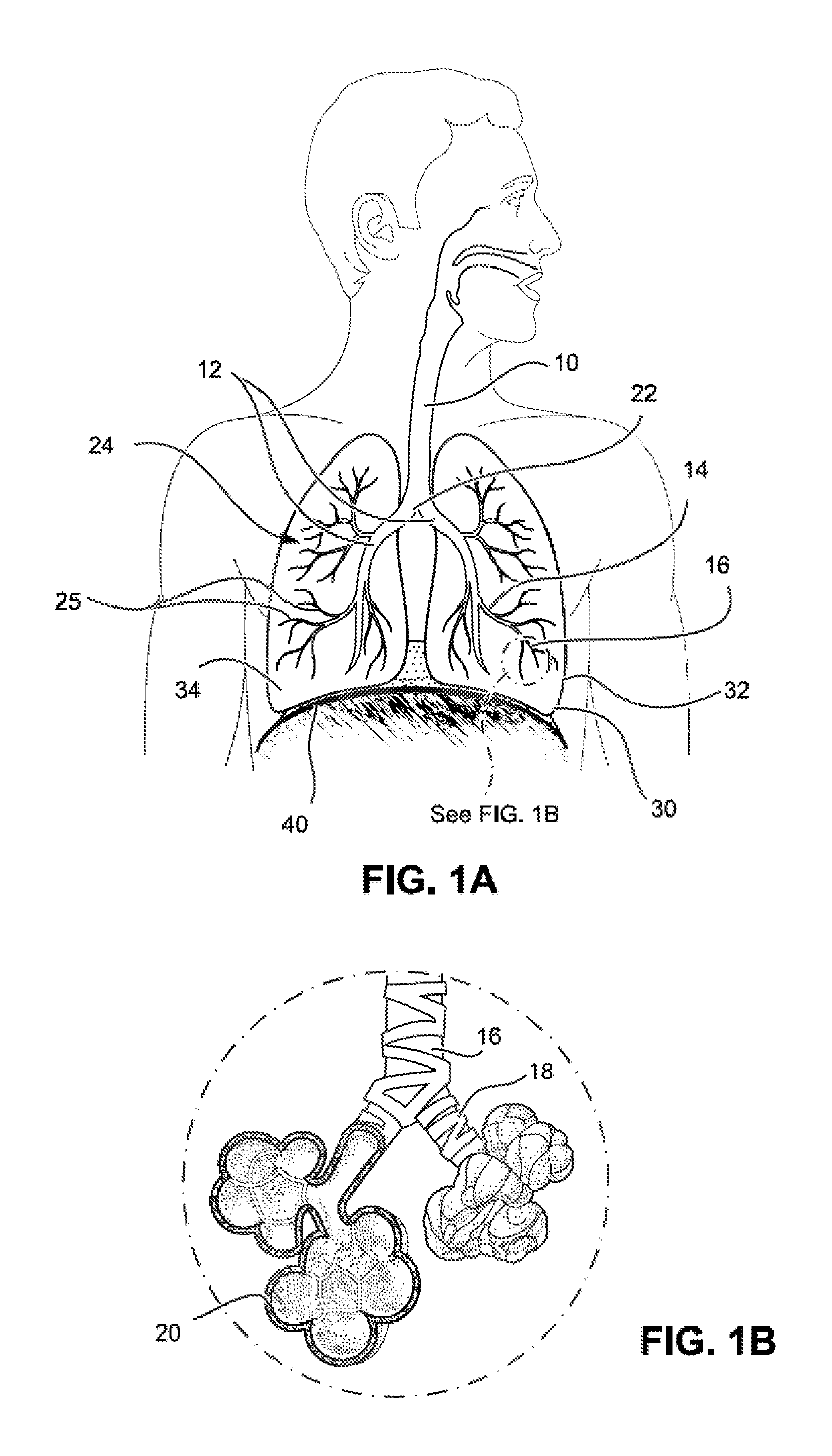
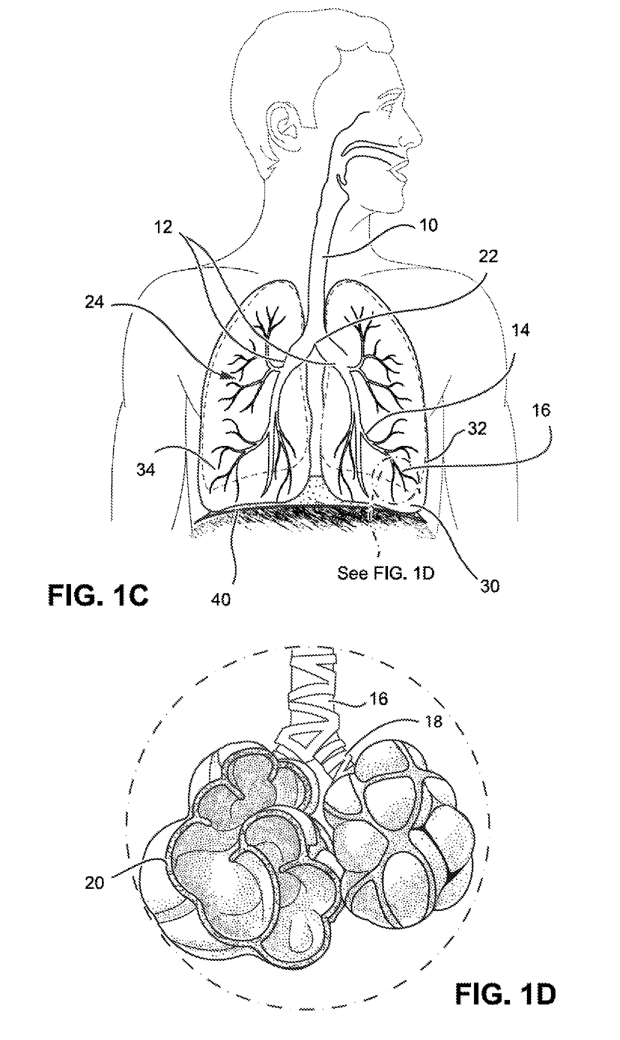
[0079]FIGS. 1A, 1B, 1C and 1D illustrate the respiratory system located primarily within the thoracic cavity. In human beings, the lungs 30 are present in pairs and are located in the pleural cavities of the thorax on either side of the heart. The lungs are separated from the abdominal cavity by the muscular thoracic diaphragm 40, which expands and contracts to facilitate breathing The lungs 30 of a typical adult human are about 25 to 30 cm long and are approximately cone shaped. A protective membrane, called the pulmonary or visceral pleura, protects the lungs and separates each lung from the parietal pleura, which covers the chest wall, by the thin layer of pleural fluid. The lungs are separated by the mediastinum, which contains the heart, trachea, esophagus and blood vessels. The lungs normally have clear anatomical divisions known as lobes. The right lung 34 is divided into three lobes called superior, middle and inferior lobes, by the oblique and horizontal fissures that are f...


PUM


Abstract

A medical device assembly including: an lung reduction device including a vertex, a first arm having an end connected to the vertex, and a second arm having an end connected to vertex, wherein the first and second arms extend into a respective one of airway branches in the lung and the vertex seats upstream of a bifurcation of the airway branches, wherein the first and second arms apply a bias force to the airway branches and thereby reduce a section of the lung near the airway branches; a bronchoscope including a channel housing the lung reduction device and having an opening to the channel through which the lung reduction device is deployed, and a pusher device associated with the bronchoscope and adapted to push the lung reduction device from the working channel to advance the first and second arms into the airway branches.

Description

FIELD OF THE INVENTION[0001]The field of the invention is lung reduction devices used to treat chronic obstructive pulmonary disease (COPD). In particular, the invention relates to lung reduction devices configured to be delivered through the airway to the lung with minimally invasive techniques.BACKGROUND OF THE INVENTION[0002]COPD is a lung disease that makes it hard to breathe. COPD can cause coughing that produces large amounts of phlegm or mucus, wheezing, shortness of breath, chest tightness, and other symptoms. Cigarette smoking is the leading cause of COPD, but long-term exposure to other lung irritants, such as air pollution, chemical fumes, or dust, may contribute to COPD. COPD is a progressive disease which gets worse over time, such as over the course of several years.[0003]To understand COPD, it helps to understand how the lungs work. Air drawn in through the nose or mouth when drawing breath goes down the windpipe into tubes in the lungs called bronchi or airways. With...

Claims


Application Information

Patent Timeline
Patent Type & Authority Applications(United States)
IPC IPC(8): A61B17/12A61B17/128A61B1/267A61F2/04
CPCA61B2017/00809A61F2002/043A61B17/1285A61B2017/0034A61F2/04A61B17/12104A61B2017/12054A61B2017/22038A61B17/12131A61B1/2676A61B17/12031A61B17/0643A61B17/1227A61B2017/00296
Inventor GELFAND, MARKWONG, ANTHONYRIOUX, ROBERT F.ENGELMAN, ZOAR
Owner EOLO MEDICAL INC
Why Eureka
Try Eureka