Methods for diagnosis, differentiation and monitoring using urine proteins as markers in iga nephropathy

a technology of urine protein and nephropathy, which is applied in the field of diagnostic methods of iga nephropathy, can solve the problems of empirics, limited treatment options, and burden on society, and achieve the effects of rapid and ultrasensitive quantitative detection, rapid characterization of proteins, and improved sensitivity and sensitivity

Active Publication Date: 2019-11-07
WARSZAWSKI UNIWERSYTET MEDYCZNY +1
View PDF4 Cites 0 Cited by
  • Summary
  • Abstract
  • Description
  • Claims
  • Application Information

AI Technical Summary

Benefits of technology

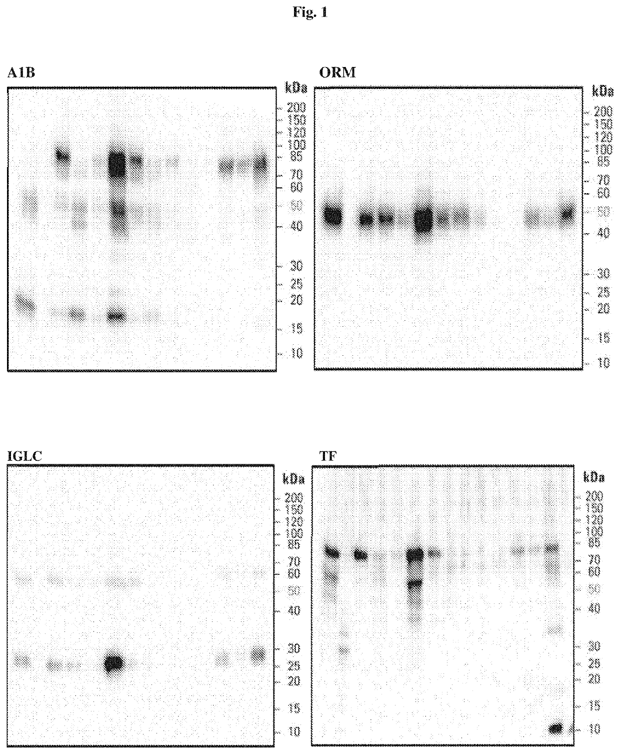
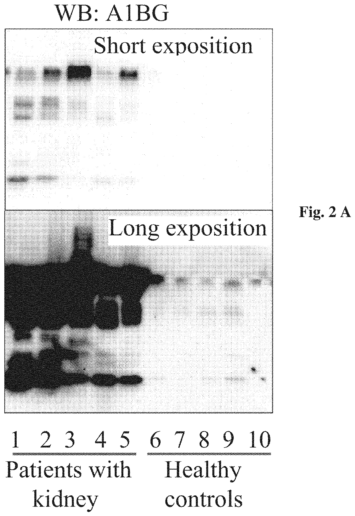
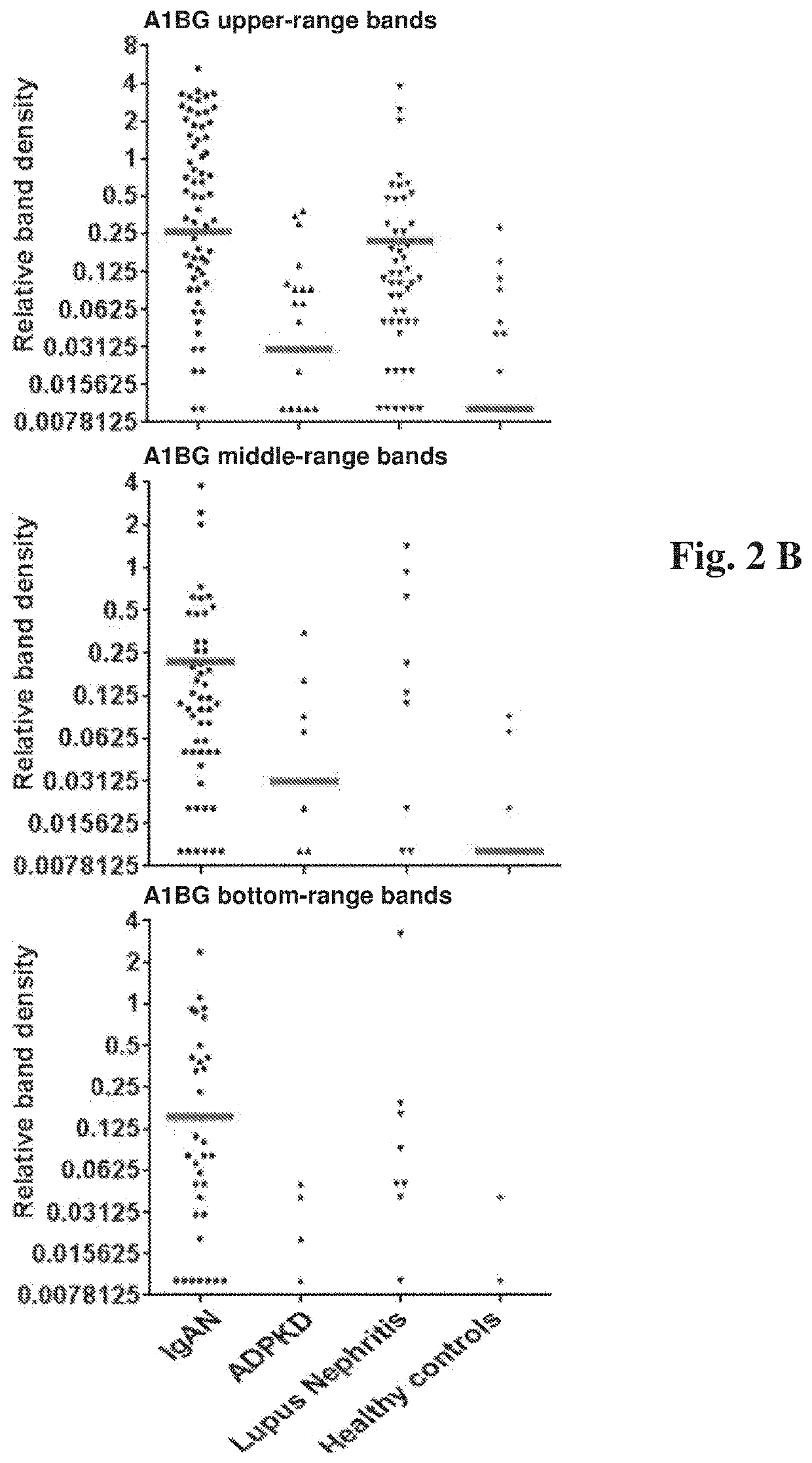
[0013]The above indicated markers are unique for IgAN and have been selected based on analysis of urine samples from patients suffering from IgAN, ADPKD, LN and healthy controls. It is herein disclosed that the levels of each protein and their non-full-length fragments, i.e. marker proteins truncated on one or both sides of the amino acid sequence of the complete protein, and / or the combination thereof correlate with the disease type, thus allowing for detection of IgAN in a patient, its differentiation from renal diseases having different etiology, and monitoring of the IgAN patient response to a treatment.
[0022]Ultrasensitive microfluidic solid-phase ELISA was reported and described, for example, in Lab Chip 2013; 13(21), 4190-4197. This method is useful in rapid and ultrasensitive quantitative detection of low abundance proteins. The microwell-based solid-phase ELISA strategy provides an expandable platform for developing the next-generation microfluidic immunoassay systems that integrate and automate digital and analog measurements to further improve the sensitivity, dynamic ranges, and reproducibility of proteomic analysis.
[0023]The other method, Microfluidic Electrophoresis Assays for Rapid Characterization of Protein, was characterized and discussed in Science / AAAS audio webinar (14 Nov. 2012) by Dr. Joey Studts from Boehringer Ingelheim in Germany, Dr. Timothy Blanc from ImClone Systems in Branchburg, N.J., and Dr. Bahram Fathollahi from PerkinElmer in San Francisco, Calif. What was discussed there concerned the application of high throughput microfluidic technologies to the analysis of biotherapeutic proteins. These microfluidic-based assays provide a good solution because they address the limitations of SDS-PAGE, as well as other separation assays that depend on conventional capillary electrophoresis in particularly analysis time, which can be reduced to a minute or less per sample. Advantages include miniaturization, integration, and automation, which enable labs to perform experiments at a rapid turnaround time, thus faster analytical analysis to reduce time and expense in the process development.
[0025]The above described analysis method makes it possible to determine marker patterns useful in monitoring of a response of a patient to a treatment for IgAN and differentiation of IgAN from other chronic kidney diseases. The solution of the invention may eliminate the need to perform biopsy to confirm diagnosis of IgAN.

Problems solved by technology

CKD represents an increasing worldwide public health problem, which causes an extensive socio-economic burden for the society.
Therefore, the treatment options are presently limited and empirics-based.

Method used


Image

  • Methods for diagnosis, differentiation and monitoring using urine proteins as markers in iga nephropathy
  • Methods for diagnosis, differentiation and monitoring using urine proteins as markers in iga nephropathy
  • Methods for diagnosis, differentiation and monitoring using urine proteins as markers in iga nephropathy

Examples


example 1

Discovery Phase

[0039]The methodological approach of the discovery phase has been described in Mucha et al. Below, the most pertinent information is listed.

Patients Characteristics

Groups of Patients

[0040]The study included 30 patients with IgAN and 30 healthy age- and sex-matched volunteers serving as controls. Demographic and clinical data of both groups are presented in Mucha, et al. (Supplementary material online, Table S1). Briefly, patients with biopsy-proven IgAN at different stages of chronic kidney disease (CKD) and older than 18 years were included. The inclusion criteria for the control group were as follows: age older than 18 years and absence of any kidney disease or other chronic diseases requiring treatment. The exclusion criteria for both groups included: active infection, history of malignancy, previous organ transplantation, or current pregnancy. To estimate GFR, we used the Chronic Kidney Disease Epidemiology Collaboration equations, which are the most accurate, hav...

example 2

Validation Phase

[0046]The primary difference between the discovery and validation phases is the transition from the assessment of the pooled urine samples (i.e. the discovery phase) to the individual evaluation of each protein in a given patient or a healthy person and a direct correlation of these results with the known clinical parameters in each case (i.e. the validation phase).

Patient Characteristics

[0047]The study included 133 renal disease patients and 19 healthy controls. Renal disease included IgAN (77 cases), ADPKD (29) and LN (27).

Sample Collection

[0048]Urinary samples were collected according to the protocol standardized in the Transplantation Institute, Medical University of Warsaw.


PUM

PropertyMeasurementUnit
weightaaaaaaaaaa
molecular weightaaaaaaaaaa

Abstract

The present invention relates to use of markers selected from a group consisting of alpha-IB-glycoprotein (A1BG), alpha-1-acid glycoprotein 1 (ORM1), Ig lambda-2 chain C regions (IGLC2) and serotransferrin (TF) in methods for diagnosis, monitoring and differentiation of IgA nephropathy. In addition corresponding diagnostic kits are provided.

Description

[0001]The Sequence Listing in ASCII text file format of 34,922 bytes in size, created on Dec. 18, 2018, with the file name “2018-12-21SequenceListing_MUCHA1,” filed in the U.S. Patent and Trademark Office on even date herewith, is hereby incorporated herein by reference.TECHNICAL FIELD[0002]The application relates the field of diagnostic methods of IgA nephropathy. The present invention is directed to methods for diagnosis, differentiation and monitoring using urine proteins as markers in IgA nephropathy. In addition corresponding diagnostic kits are provided.BACKGROUND OF THE INVENTION[0003]IgA nephropathy (IgAN) is the most common primary glomerulonephritis worldwide that may lead to a chronic kidney disease (CKD). CKD represents an increasing worldwide public health problem, which causes an extensive socio-economic burden for the society. The prevalence of CKD is up to 14.2% in the USA, 10.2% in Norway, and 11.9% in Poland. It is estimated that over 4 million people in Poland suf...

Claims


Application Information

Patent Timeline
Patent Type & Authority Applications(United States)
IPC IPC(8): G01N33/68G01N33/493
CPCG01N33/6827G01N33/6848G01N33/493G01N2800/347G01N33/6857G01N33/6893G01N2333/473G01N2333/79G01N2800/52
Inventor MUCHA, KRZYSZTOFPACZEK, LESZEKZAGOZDZON, RADOSLAWFORONCEWICZ, BARTOSZDADLEZ, MICHALBAKUN, MAGDALENAPIWOWARSKI, JANPILZYS, TOMASMARCINKOWSKI, MICHALGARBICZ, DAMIANGRZESIUK, ELZBIETAFLORCZAK, MICHAL
Owner WARSZAWSKI UNIWERSYTET MEDYCZNY
Why Eureka
Try Eureka