Medical image processing device and medical image photographing system

Active Publication Date: 2020-01-23
KONICA MINOLTA INC
View PDF0 Cites 0 Cited by
  • Summary
  • Abstract
  • Description
  • Claims
  • Application Information

AI Technical Summary

Benefits of technology

[0007]It is an object of the present disclosure to easily ac

Problems solved by technology

However, it is not possible with conventional bone factor measurement methods to easily acquire the

Method used


Image

  • Medical image processing device and medical image photographing system
  • Medical image processing device and medical image photographing system
  • Medical image processing device and medical image photographing system

Examples


Embodiment Construction

[0042]Hereinafter, one or more embodiments of the present disclosure will be described with reference to the drawings. However, the scope of the disclosure is not limited to the disclosed embodiments. The embodiments according to the present disclosure will be described with reference to the accompanying drawings.

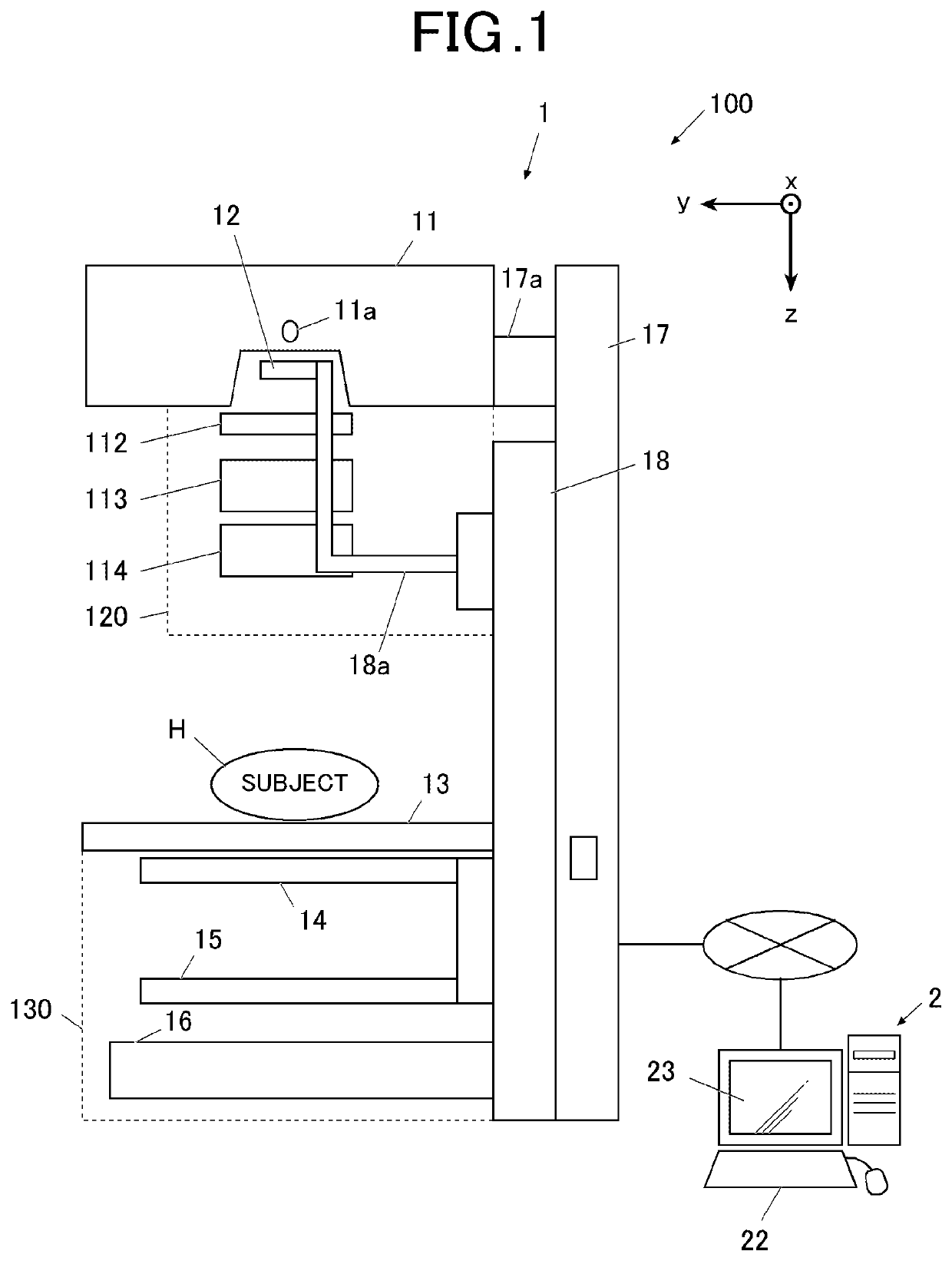
[0043]First, a device configuration according to the embodiment will be described with reference to FIG. 1 to FIG. 4. FIG. 1 is a schematic diagram illustrating an X-ray photographing system 100 according to the embodiment.

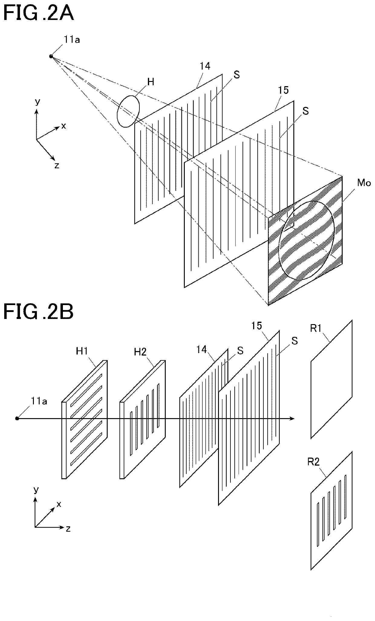
[0044]As illustrated in FIG. 1, in the embodiment, the X-ray photographing system 100 as a medical image photographing system is used. The X-ray photographing system 100 includes an X-ray Talbot photographing device 1 as a medical image photographing device, and an image processing device 2 as a medical image processing device. A same body part of a subject H is photographed for a plurality of times by using the X-ray Talbot photographing device 1 while c...


PUM


Abstract

Disclosed is a way to provide a medical image processing device that may include a hardware processor that calculates at least one of trabecular connectivity, trabecular width, trabecular number, mineralization degree, osteoid volume, cortical width, and cortical porosity as a bone characteristic indicator of a subject from reconstructed image data generated from moiré image data acquired by photographing the subject.

Description

CROSS REFERENCE TO RELATED APPLICATIONS[0001]This application claims priority to Japanese Patent Application No. 2018-136288, filed on Jul. 20, 2018, which is incorporated herein by reference in its entirety.BACKGROUNDTechnological Field[0002]The present disclosure relates to a medical image processing device and a medical image photographing system.Description of the Related Art[0003]As an X-ray photographing device, conventionally known is an X-ray photographing device (referred to as an X-ray Talbot photographing device hereinafter) using a Talbot interferometer or a Talbot-Lau interferometer having one-dimensional grating. Further, through reconstructing moiré images (moiré fringe images) photographed by the X-ray Talbot photographing device, it is possible to acquire at least three kinds of high-definition reconstructed images such as an absorption image, a differential phase image, and a small-angle scattering image.[0004]Further, also known is a medical image system that gene...

Claims


Application Information

Patent Timeline
IPC IPC(8): A61B6/00G06T7/00
CPCA61B6/483A61B6/463G06T7/0012A61B6/5217A61B6/5235G06T11/00A61B5/4509A61B6/4291A61B6/505G16H30/40A61B6/484A61B6/4035A61B6/5205A61B6/482A61B6/5282A61B6/469G06T2207/10116G06T2207/30008G16H50/30A61B5/4504A61B5/004
Inventor SHINDEN, YUKOHOSHINO, YOSHIHIDE
Owner KONICA MINOLTA INC
Why Eureka
Try Eureka