Functional measurements in echocardiography

a functional measurement and echocardiography technology, applied in the field of functional measurement in echocardiography, can solve the problems of poor inter- and intravariability of methods, poor temporal resolution and ad-hoc setup, and other problems, to achieve the effect of reducing potential burst nois

Inactive Publication Date: 2020-03-05
NORWEGIAN UNIVERSITY OF SCIENCE AND TECHNOLOGY (NTNU)
View PDF0 Cites 3 Cited by
  • Summary
  • Abstract
  • Description
  • Claims
  • Application Information

AI Technical Summary

Benefits of technology

[0018]At step (iv) measurements are fused via state estimation (for example via a Kalman filter) employed on every point-velocity component in the measurement kernel (i.e. all data...

Problems solved by technology

Despite being a central part of standard protocol examinations at the outpatient clinic, the methods tend to have poor inter- and intravariability.
However, these methods also have several limitations.
A poor parall...

Method used


Image

  • Functional measurements in echocardiography
  • Functional measurements in echocardiography
  • Functional measurements in echocardiography

Examples


Embodiment Construction

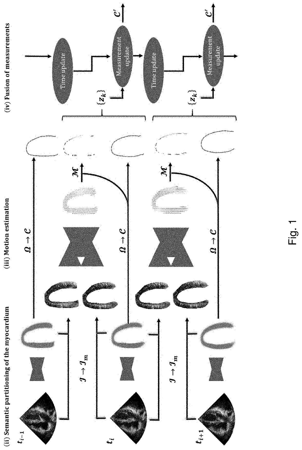
[0052]As described in further detail below, a method has been developed that enables automatic functional measurements in 2D echocardiography. The system works in an end-to-end fashion with standard cardiac ultrasound images as input, and several clinical measures, as well as motion estimates and regional masks as direct output.

[0053]The method core is comprised of five components, (i) classification of cardiac view, (ii) segmentation and semantic partitioning of the left ventricle (LV) myocardium, (iii) regional motion estimates, (iv) fusion of measurements and (v) calculation of clinical indices. An illustration of an example setup for measuring global longitudinal strain after view classification is illustrated in FIG. 1. By way of an overview, FIG. 1 shows how ultrasound images are forwarded through a segmentation network, and the resulting masks are used to extract relevant parts of the image. The masked ultrasound data is further processed through the motion estimation network...


PUM


Abstract

A method for processing echocardiography data to enable automatic functional measurements based on cardiac ultrasound images as an input, including (i) classification of the cardiac ultrasound images to ensure that relevant images are passed on to the next steps, optionally utilising a first neural network, such as a convolutional neural network, (ii) segmentation and semantic partitioning of the left ventricle (LV) myocardium to extract relevant parts of the image, optionally by using a second neural network, (iii) regional motion estimates to determine a mapping of displacements in the extracted parts of the image and to output estimated tissue motion vectors for the extracted parts of the image, optionally using a third neural network, and (iv) fusion of measurements via state estimation applied to the tissue motion vectors and thereby incorporating a temporal domain to produce data showing variation of the estimated measurements over time.

Description

TECHNICAL FIELD[0001]The invention relates to a method for processing echocardiography data in order to allow for functional measurements to be obtained automatically. The invention further provides corresponding computer programme products and systems.BACKGROUND OF THE INVENTION[0002]Recent years have shown that quantitative assessment of cardiac function has become indispensable in echocardiography. Evaluation of the hearts contractile apparatus has traditionally been limited to geometric measures such as ejection fraction (EF) and visual estimation (eyeballing) of myocardial morphophysiology. Despite being a central part of standard protocol examinations at the outpatient clinic, the methods tend to have poor inter- and intravariability. With tissue doppler imaging (TDI) and speckle tracking (ST), the quantification tools have moved beyond these measures, and enabled new methods for assessing the myocardial deformation pattern. Myocardial deformation imaging, e.g. strain and stra...

Claims


Application Information

Patent Timeline
IPC IPC(8): G06T7/00G06N3/04G16H30/40G06T7/11G06T7/20G06K9/62A61B8/08A61B8/14
CPCG06N3/0454G06T7/0012G06T2207/20084G06T7/11G06K9/6267G06T2200/24G06T7/20G06T2207/10132A61B8/14G06K9/6228G06K9/6261A61B8/0883G06T2207/30048G16H30/40G06K2209/051G06T7/0016G06V2201/031G06F18/24G06F18/211G06F18/2163G06N3/045
Inventor ØSTVIK, ANDREASSMISTAD, ERIKLØVSTAKKEN, LASSE
Owner NORWEGIAN UNIVERSITY OF SCIENCE AND TECHNOLOGY (NTNU)
Why Eureka
Try Eureka