Automated lesion detection, segmentation, and longitudinal identification

a technology of lesion detection and segmentation, applied in the field of automatic lesion detection, segmentation and longitudinal identification, can solve the problems of increasing the difficulty of synthesizing the results from the many gathered series, the difficulty of careful quantitative assessment of lung and liver lesions, and the inability to provide as much information as mri

Inactive Publication Date: 2020-03-19
ARTERYS INC
View PDF0 Cites 151 Cited by
  • Summary
  • Abstract
  • Description
  • Claims
  • Application Information

AI Technical Summary

Problems solved by technology

CT is simpler to gather and read, but it does not provide as much information as MRI.
However, there is increased difficulty associated with synthesizing the results from the many gathered series compared with reading a single CT scan.
Careful quantitative assessment of lung and liver lesions is tedious and time consuming.
However, even with training, radiologists are prone to fatigue and mistakes.
In addition, after ROIs are detected, quantitative assessment, such as calculating the volume via segmentation, requires additional time and effort.
Finding regions of interest in a volumetric image is a challenging task for both humans and computer algorithms alike.
Multiple radiologists reading the same scan often identify different regions as being cause for concern and disagree about likelihood.
However, they also have imperfect sensitivity.
However, multiple of these stages require user input (e.g., placement of seed points) and review, resulting in a slower diagnosis than a more fully-automated method.
However, while CT intensities are reproducible, lesion intensities and locations can vary greatly; this makes this algorithm highly susceptible to accidental inclusion of lesions in the segmentation of benign pulmonary structures.
HOG features do not fully characterize the lesion, as they do not consider global context, a major limitation that prevents the classifier from learning lesion shapes.
PCA limits the scope of the features found to a subset of all features available, which inherently limits the classifier to capturing only lesions that possess the retained features.
Additionally, SVMs do not scale well; given the same amount of data, deep learning models are able to train more efficiently and pick up on more subtle details, resulting in a higher accuracy upper limit.
The resulting contour incorrectly spills into the chest wall.
Although the snakes algorithm and other deformable models that rely on a shape prior are common, and although modifying its resulting contours can be significantly faster than generating contours from scratch, the snakes algorithm has several significant disadvantages.
The cost function typically awards credit when the contour overlaps edges in the image; however, there is no way to inform the algorithm that the edge detected is the one desired; e.g., there is no explicit differentiation between the edge of the ROI and blood vessels, airways, or other anatomy.
Furthermore, these algorithms are greedy.
However, gradient descent, and many similar optimization algorithms, are susceptible to getting stuck in local minima of the cost function.
Because they generally only have a few dozen tunable parameters, the algorithms do not have the capacity to represent a diverse set of possible images on which segmentation is desired.
Because of the great diversity on recorded images and the small number of tunable parameters, a snakes algorithm or deformable model can only perform well on a small subset of “well-behaved” cases.
However, the snakes algorithm cannot be adequately tuned to work on more challenging cases.

Method used


Image

  • Automated lesion detection, segmentation, and longitudinal identification
  • Automated lesion detection, segmentation, and longitudinal identification
  • Automated lesion detection, segmentation, and longitudinal identification

Examples


1st embodiment (

CT Example): Lund Augmented Workflow

[0357]GUI that Comprises Automated and Manual Tools for Chest CT Analyses

[0358]Setup

[0359]FIG. 47 shows a screenshot 4700 of an example GUI that allows for several lung CT studies to be displayed next to each other and be co-registered so that the same anatomy in the scans shows at the same time (e.g., 4702 and 4704). The image brightness and contrast may be automatically adjusted for optimal lung reading. Furthermore, this user interface can display several studies of this type at the same time in order to make it easy for the physician to compare images from the same patient over time. In both cases, the physician can scroll through studies, zoom, and move images to see the same anatomy in all of the different studies simultaneously. The system also offers manual and automated tools to level the brightness and contrast of the image based on the workflow selected.

[0360]Detection

[0361]The system is built to automatically detect and measure finding...

1st embodiment

[0409]Overview

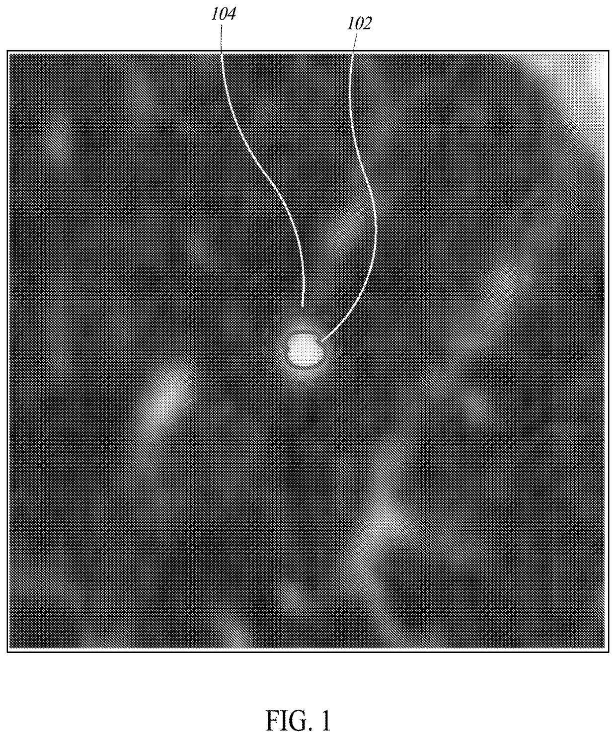
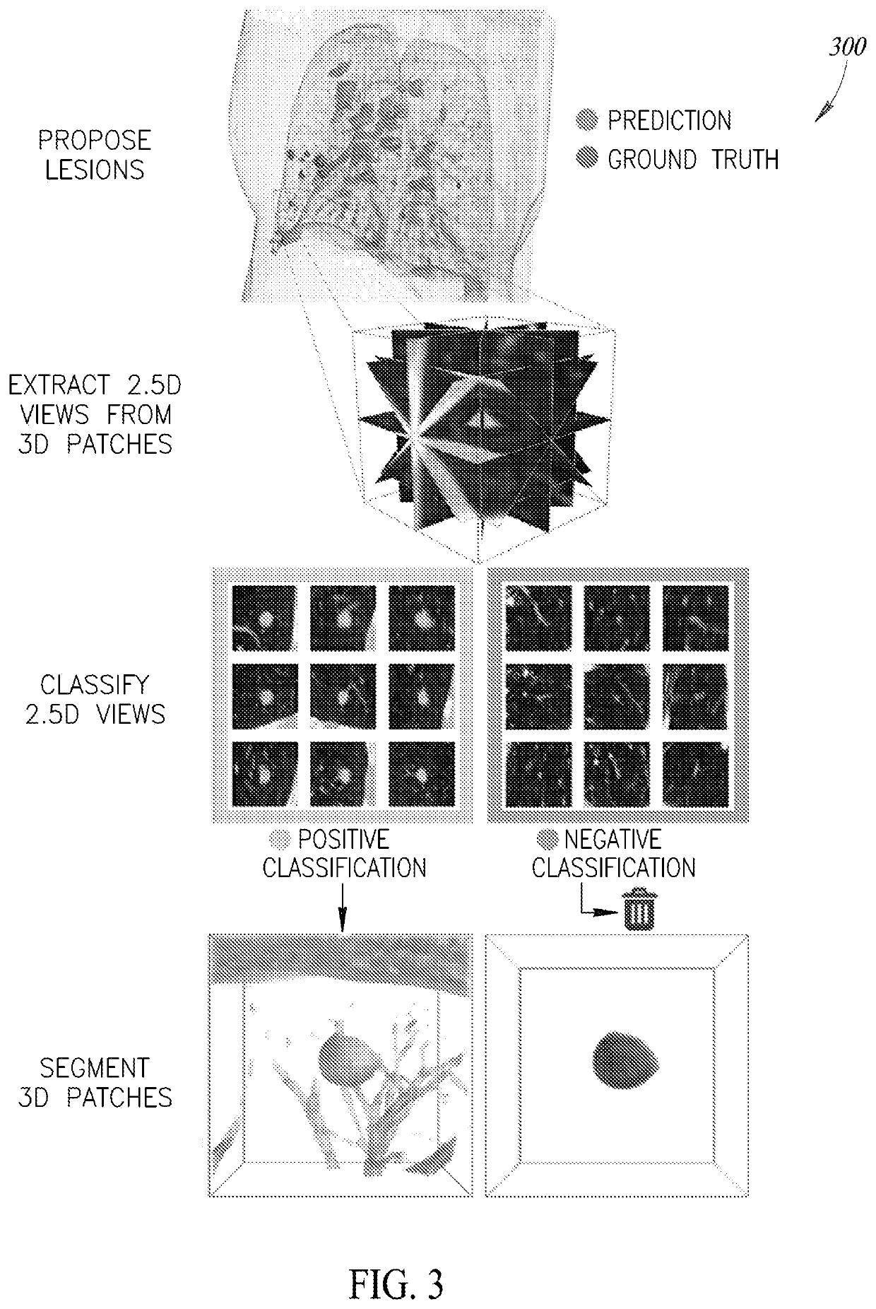
[0410]FIG. 54 is a flow diagram of a process 5400 of operating a processor-based system to store information about a pre-localized region of interest in image data and to reveal such information upon user interaction, according to one illustrated implementation. The process 5400 begins at 5402 when image data is uploaded to a processor-based system. A pre-trained algorithm for lesion localization stored in a database at 5404 is used to localize lesions in the image data at 5406. This pre-trained algorithm may include one or more machine learning algorithms, such as, but not limited to, Convolutional Neural Networks (CNNs). In at least one implementation of the current disclosure, two unique CNNs are joined end to end; the first CNN proposes locations of potential lesions with a focus on high sensitivity, and the second CNN sorts through these proposed lesions and discards results determined to be false positives.

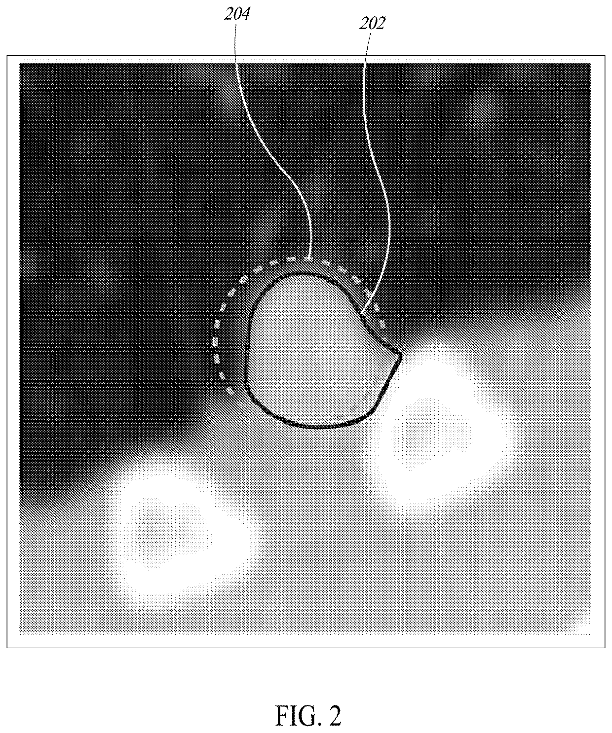
[0411]A pre-trained CNN model for segmentation of lesions...


PUM


Abstract

Computed Tomography (CT) and Magnetic Resonance Imaging (MRI) are commonly used to assess patients with known or suspected pathologies of the lungs and liver. In particular, identification and quantification of possibly malignant regions identified in these high-resolution images is essential for accurate and timely diagnosis. However, careful quantitative assessment of lung and liver lesions is tedious and time consuming. This disclosure describes an automated end-to-end pipeline for accurate lesion detection and segmentation.

Description

OVERVIEW[0001]Various implementations of the present disclosure are discussed herein below. For readability, the implementations are provided under separate headings. In particular, the following top-level headings are provided for the various implementations: Automated Lesion Detection, Segmentation, and Longitudinal Identification; Content Based Image Retrieval for Lesion Analysis; Three Dimensional Voxel Segmentation Tool; Systems and Methods for Interaction with Medical Image Data; Automated Three Dimensional Lesion Segmentation; Autonomous Detection of Medical Study Types; Patient Outcomes Prediction System; and Co-registration. It should be appreciated that the discussion relating to one or more implementations may be applicable to one or more other implementations. Further, features of each of the various implementations discussed herein may be combined with one or more other implementations to provide additional implementations.A. Automated Lesion Detection, Segmentation, an...

Claims


Application Information

Patent Timeline
Patent Type & Authority Applications(United States)
IPC IPC(8): A61B5/00A61B5/055A61B6/03A61B6/00G06N3/04G06N3/08G06T7/00G06V10/764
CPCG06T2207/10088G06T2207/10081G06N3/0454G06N3/084G06N3/082G06T7/0016G06T2207/30056G06T2207/20084A61B6/5217A61B5/055A61B6/563A61B6/032G06T2207/30064G06T2207/20081A61B5/7267G06T2207/30096A61B5/7264G06N10/00G16H50/30G06T7/30G06V2201/031G06V10/82G06V10/764G06N3/045G06F18/24143
Inventor TAERUM, TORIN ARNILAU, HOK KANSALL, SEANLE, MATTHIEUAXERIO-CILIES, JOHNGOLDEN, DANIEL IRVINGLIEMAN-SIFRY, JESSEJUGDEV, TRISTAN
Owner ARTERYS INC
Why Eureka
Try Eureka