Fabp4 as a therapeutic target in skin diseases
- Summary
- Abstract
- Description
- Claims
- Application Information
AI Technical Summary
Benefits of technology
Problems solved by technology
Method used
Image
Examples
example 1
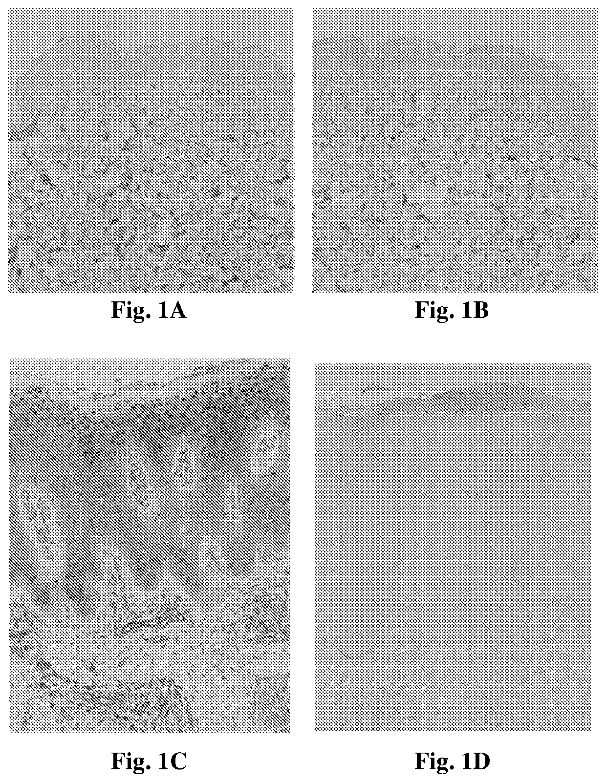
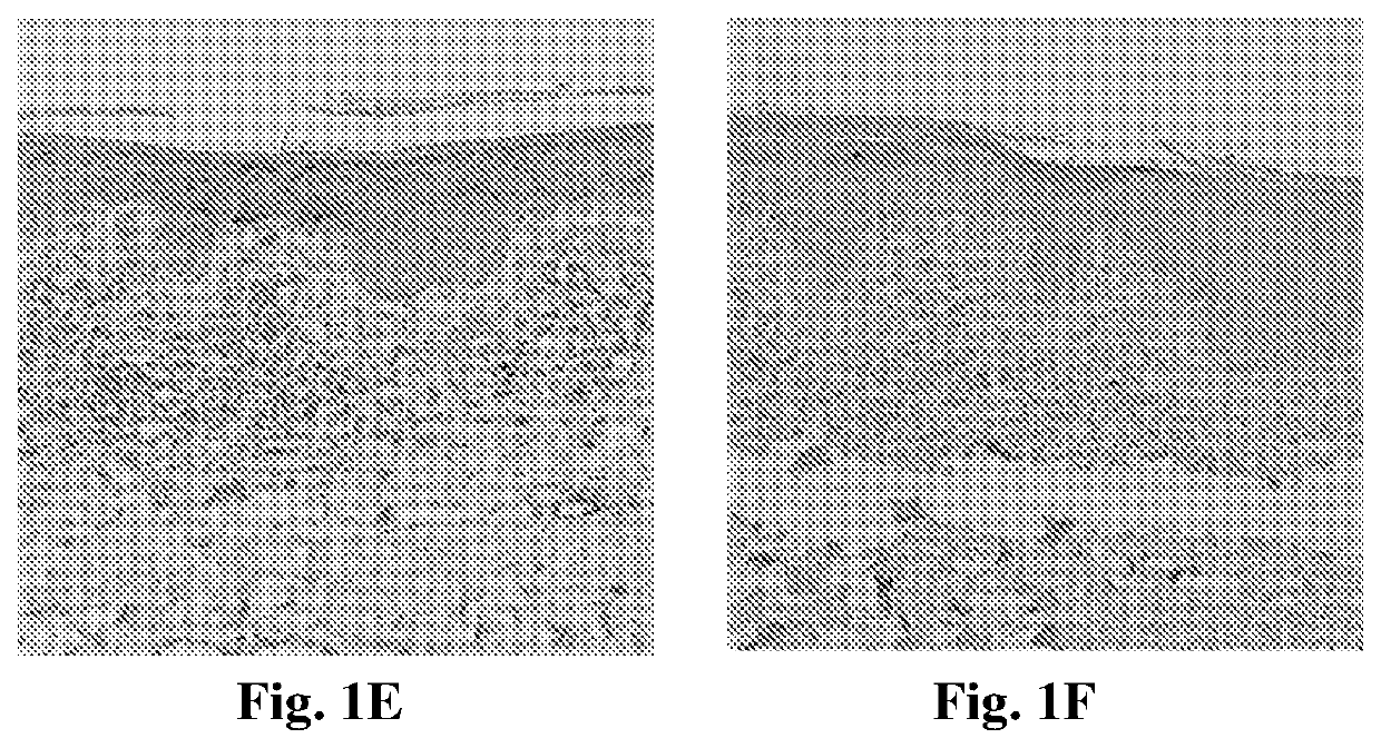
Tissue Expression Analysis of FABP4, FABPS, and PPARγ in Human Psoriatic Skin Lesions
[0117]Punch biopsies (4 mm in diameter) were obtained from disease-involved skin of patients with psoriasis (n=10). Biopsies were further obtained from involved skin of patients with chronic dermatitis (n=5). Normal skin was obtained from patients after surgical reduction of redundant skin (n=10). The tissues were fixed in formalin and embedded in paraffin. For histopathological examination with light microscopy, the sections were stained with hematoxylin and eosin (H&E) and observed by a pathologist who confirmed the diagnosis. In addition, for immunohistochemical analysis, the sections were stained with the following antibodies: FABP4 (Rabbit polyclonal anti FABP4 antibody, PAB12276, Abnova), FABPS (Rabbit polyclonal anti FABPS antibody, SC-50379, Santa Crus) and PPARγ (mouse monoclonal anti-PPARγ antibody, E-8, Santa Crus), all diluted 1:50. The paraffin-embedded and cryopreserved tissues were pr...
example 2
Tissue Expression Analysis of FABP4 in Human Cutaneous T Cell Lymphoma (CTCL) Skin Lesions
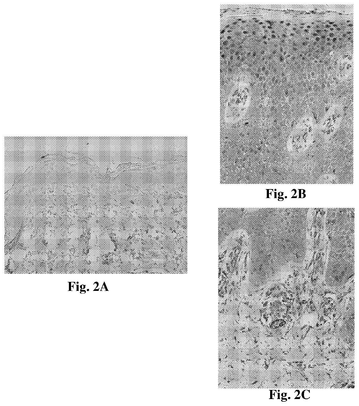
[0123]CTCL is a group of neoplasms of skin-homing T cells. Mycosis fungoides (MF) represents the most common type of CTCL and accounts for approximately 50% of all primary cutaneous lymphomas. Although malignant in nature, MF has a protracted clinical course with inflammatory, dermatitis-like presentation [8].
[0124]The expression of FABP4 among patients with MF was studied by performing an immunohistochemical analysis of lesional skin. Samples from 5 patients with MF were tested and compared to normal human skin. Punch biopsies (6 mm in diameter) were obtained from patients with MF, from disease-involved skin. Normal skin was obtained from patients after surgical reduction of redundant skin. Tissue sections were processed and stained with anti-FABP4 antibody as described in Example 1.
[0125]High expression levels of FABP4 were observed in MF lesions compared to normal skin with all tested patien...
example 3
Overexpressing FABP4 in Primary Murine Keratinocytes
[0128]The introduction of FABP4 into keratinocytes was suggested to create a hyperproliferative state with impaired differentiation as seen in psoriasis. Differentiation was assessed by measuring the expression of two keratinocyte differentiation markers K1 and K5. K5 is a keratin whose level does not change during keratinocyte differentiation and therefore serves as a loading control, while K1 is induced during normal keratinocyte differentiation.
[0129]For assessing the effect of FABP4 overexpression in mouse primary keratinocytes, cells were infected with a lentiviral vector construct containing the FABP4 gene. The FABP4 lentiviral vector, named FABP4-T2A-EGFP, was constructed with the FABP4 gene under the CMV promotor, and contained the green fluorescence protein (GFP) gene as a reporter of expression. The presence of a T2A residue at the N-terminus of the FABP4 protein precludes FABP4 detection by anti-FABP4 commercial antibodi...
PUM
| Property | Measurement | Unit |
|---|---|---|
| Mass | aaaaa | aaaaa |
| Mass | aaaaa | aaaaa |
| Molecular weight | aaaaa | aaaaa |
Abstract
Description
Claims
Application Information
 - R&D
- Intellectual Property
- Life Sciences
- Materials
- Tech Scout
- Unparalleled Data Quality
- Higher Quality Content
- 60% Fewer Hallucinations
Browse by: Latest US Patents, China's latest patents, Technical Efficacy Thesaurus, Application Domain, Technology Topic, Popular Technical Reports.
© 2025 PatSnap. All rights reserved.Legal|Privacy policy|Modern Slavery Act Transparency Statement|Sitemap|About US| Contact US: help@patsnap.com


