Extracellular vesicle markers for stable angina and unstable angina

a technology of extracellular vesicle markers and stable angina, applied in the field of medicine, can solve the problems of ischemic heart disease, a major cause of morbidity and mortality worldwide, atypical symptoms, and shortness of breath

Inactive Publication Date: 2020-05-28
UMC UTRECHT HLDG BV
View PDF0 Cites 1 Cited by
  • Summary
  • Abstract
  • Description
  • Claims
  • Application Information

AI Technical Summary

Benefits of technology

The present invention provides a method for identifying subjects suffering from or at risk of ischemic heart disease using a plasma sample. The method involves measuring the concentration of certain protein markers in different plasma fractions, such as LDL, HDL, REX, and TEX. By comparing these concentrations to a reference value, a statistically significant difference indicates a subject with the disease. The method can provide a reliable and accurate way to identify ischemic heart disease, even at a early stage.

Problems solved by technology

Ischemic heart disease is a major cause of morbidity and mortality worldwide.
Stable coronary artery disease can also cause atypical symptoms such as shortness of breath or reduced exercise tolerance.
Because of the variety in clinical presentation and a broad differential diagnosis (such as musculoskeletal or psychological problems, pulmonary embolism, pneumonia, pneumothorax, pericarditis), the diagnosis of stable angina is notoriously challenging.
Such imaging tests have a sensitivity and specificity of around 85%, and are time consuming and expensive.
The diagnostic work-up is inefficient and expensive.
Rapid diagnosis of unstable angina is essential, since it is associated with a high risk of adverse cardiac events; more than in the case of stable angina.
As for stable angina, the diagnosis for unstable angina is similarly challenging.
No rapidly determinable and reliable diagnostic markers are available that positively identify the unstable angina patient, or that will on the other hand, exclude unstable angina.
Although these markers have some prognostic value, none of them appear to have sufficient diagnostic power to discriminate patients with stable and unstable angina (Tsaknis et al.
However, these miRNA have thus far not been validated.
Next to this, detection of miRNA is technically challenging requiring cDNA synthesis and qPCR which limits their application in an acute setting (such as with unstable angina) or in GP settings.

Method used


Image

  • Extracellular vesicle markers for stable angina and unstable angina
  • Extracellular vesicle markers for stable angina and unstable angina
  • Extracellular vesicle markers for stable angina and unstable angina

Examples


example 1

Study on Minerva Unstable Angina Cohort

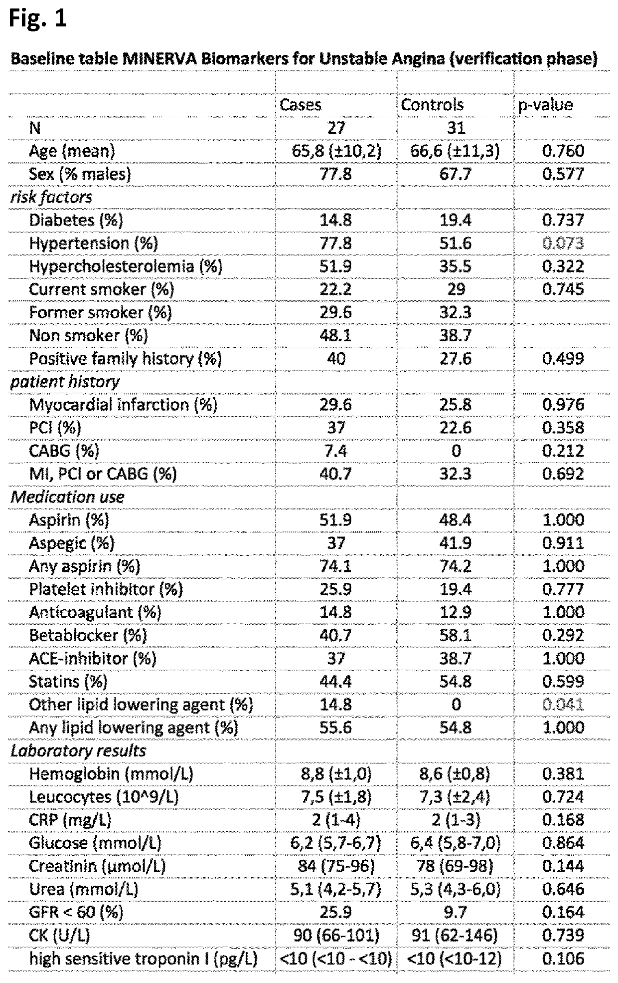
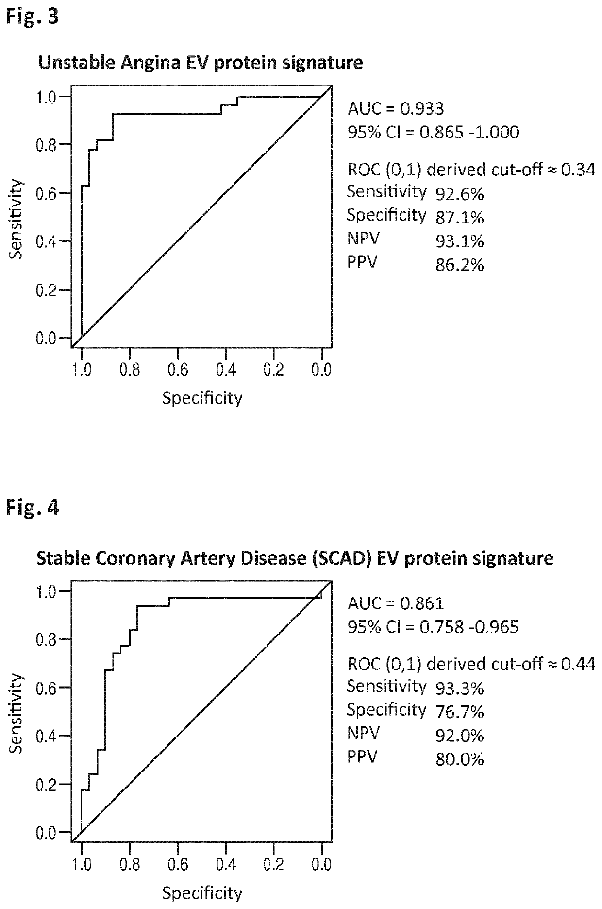
[0183]The MINERVA study (abbreviation for determination of Microvesicle content IN the Emergency Room: diagnostic Value for Acute coronary syndromes) is a single center, prospective cohort study of patients presenting to the emergency department within 24 hours of the onset of chest pain suggestive of acute coronary syndrome (ACS). Between January 2012 and June 2014, over 2000 consecutive patients were enrolled in the Meander Medical Center Amersfoort, the Netherlands, which is a large regional teaching hospital providing healthcare for a population of ˜300,000 patients. Patients younger than 18 years, patients who were unable or unwilling to give their informed consent and patients with a clear-cut ST-segment elevation MI (STEMI) were not included in this study. All patients were evaluated and managed according to the international guidelines. Along with the routine clinical laboratory measurements directly upon presentation, additional 3×10 c...

example 2

Stable Angina Cohort

[0202]The MYOMARKER stable coronary artery disease cohort (abbreviation for MYOcardial ischemia detection by circulating bioMARKERs) is a single center, prospective cohort study of patients, who are evaluated in the Meander Medical Center Cardiology outpatient clinic for (recent onset) suspected symptomatic coronary artery disease, undergoing radionuclide myocardial perfusion imaging (rMPI) as indicated by their own cardiologist. Ahead of rMPI, venous blood (6×10 cc) is obtained from the peripheral intravenous cannula, which is inserted as part of standard preparation for rMPI. The plasma component is frozen and stored at −80° C. within 1 hour after sample collection. All patients are evaluated and managed according to the international guidelines, by their own cardiologist. Patient enrollment started in August 2014 and continues until at least 1250 patients are participating in the study. At the filing of the present application (November 2015) over 800 patients...

example 3

ion of Proteins Associated with Extracellular Vesicles

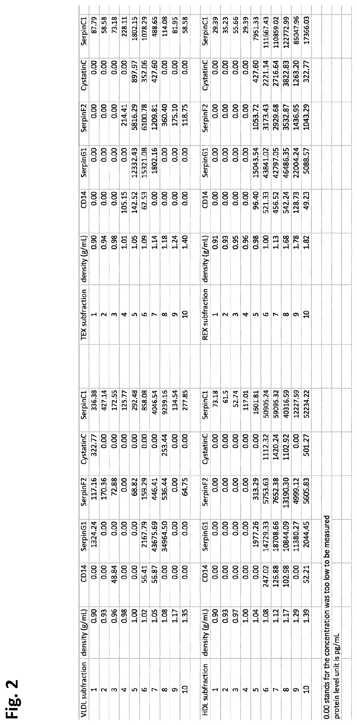
[0207]In order to determine if the five proteins selected were present in or on extracellular vesicles (EV), density gradient ultracentrifugation was performed. VLDL-EV, HDL-EV, REX-EV and TEX-EV fractions were isolated from 8 mL plasma as mentioned under example 1. Each of the precipitates (LDL, HDL, REX, TEX) was re-suspended in 500 μl PBS and used for the density gradient centrifugation. In order to make the density gradient buffer, 5 solutions containing different concentrations of OptiPrep™ medium (OptiPrep, Axis-shiled #1114542) were prepared with 10×PBS pH 7.4 (Ambion®, Life Technologies # AM9265) and ddH2O. The working solutions comprised 5%, 10%, 20%, 30% and 40% OptiPrep and 1×PBS respectively. The solutions were added and overlaid into the ultracentrifuge tubes (Beckman Coulter #344059) sequentially from the highest density one (with the highest concentration of OptiPrep) to the lowest density one (with the lowest conc...


PUM


Abstract

The present invention relates to the determination of protein markers associated with extracellular vesicles present in sub-fractions of plasma samples taken from people that experience chest pain and are suspected to experience ischemic heart disease. The invention relates to the use of the markers in the identification of subjects suffering from, or at risk of suffering from, an ischemic heart disease, in particular stable angina and unstable angina. Especially preferred markers are SerpinC1, SerpinG1, CD14, Cystatin C and SerpinF2.

Description

FIELD OF THE INVENTION[0001]The invention relates to the field of medicine. More in particular it relates to a method for identifying a subject suffering from, or being at risk of suffering from, an ischemic heart disease by measuring the concentration or a value related thereto of one or more protein markers in one or more blood plasma fractions. The protein markers are especially suitable for identifying a subject suffering from, or being at risk of suffering from unstable and stable angina, and equivalents thereof.BACKGROUND ART[0002]Ischemic heart disease is a major cause of morbidity and mortality worldwide. Although therapy for ischemic heart disease has greatly improved and mortality has gradually declined in industrialized countries during the last decades, mortality from ischemic heart disease is still rising in other parts of the world such as Africa and parts of Asia (Mathers and Loncar. Projections of global mortality and burden of disease from 2002 to 2030. PLoS Med. 20...

Claims


Application Information

Patent Timeline
IPC IPC(8): G01N33/68
CPCG01N2333/8121G01N2333/8139G01N33/6893G01N2800/324G01N2333/70596G01N33/92
Inventor DE KLEIJN, DOMINICUS PASCHALIS VICTORTIMMERS, LEONARDUS
Owner UMC UTRECHT HLDG BV
Why Eureka
Try Eureka