
Small tube tissue biopsy

a biopsy and small tube technology, applied in medical science, ovulation diagnostics, surgery, etc., can solve the problems of limiting treatment options, increasing the risk of surgical procedures, and difficult early detection of ovarian cancer, so as to avoid the risk of vessel rupture and minimal irritation of the vessel

Pending Publication Date: 2020-10-15
GW MEDICAL LLC
View PDF3 Cites 2 Cited by
  • Summary
  • Abstract
  • Description
  • Claims
  • Application Information

AI Technical Summary

Benefits of technology

The patent describes biopsy apparatuses that can safely remove biopsy samples from within a small-diameter lumen of a body without risking rupture of the vessel and with minimal irritation to the vessel. These apparatuses include a small-tube biopsy device that can be gently removed from within the vessel wall using a high-density knit, high-weave, or high-density wire. The device has a stable structure formed by applying a compressive force on the braided textile to prevent buckling or bending. The apparatuses can be used in a variety of procedures and may be housed within a delivery catheter for protection and easy removal from the body.

Problems solved by technology

Early detection of ovarian cancer may be difficult due to a lack of effective screening tests, such that ovarian cancer may not be diagnosed until the disease has reached advanced stages, limiting treatment options.
Any surgical procedure increases a risk to the patient, including but not limited to experiencing an adverse reaction, and / or requiring significant recovery time.
Additionally, an ovary biopsy may expose the patient to additional risk of potentially spreading diseased (e.g., cancerous) cells.

Method used


Image

  • Small tube tissue biopsy
  • Small tube tissue biopsy
  • Small tube tissue biopsy

Examples


Embodiment Construction

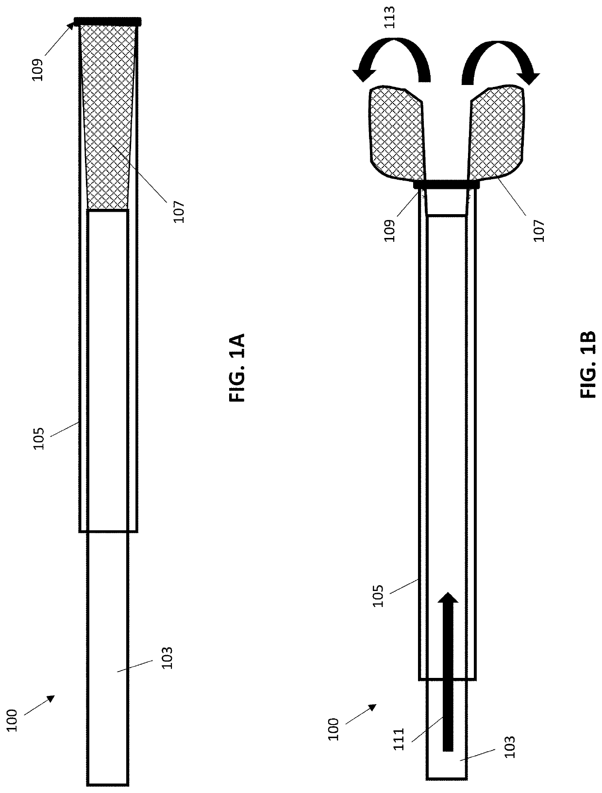
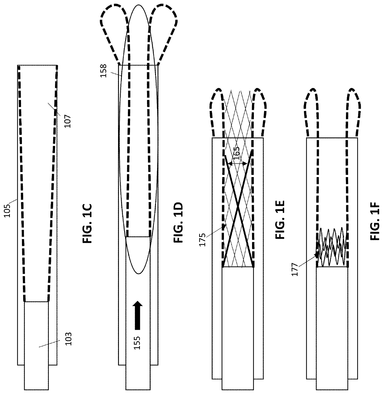
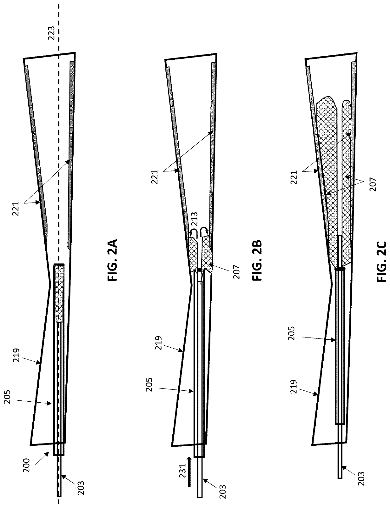
[0070]Described herein are method and apparatuses (e.g., devices and systems) for taking a biopsy from the inside of a small tube without rupturing or dissecting the small tube. The methods and apparatuses described herein may be used for any body lumen, including in particular small-diameter body lumen, such as the fallopian tubes, urethra, etc.

[0071]In general, these devices may include a first end of a biopsy collector (e.g., biopsy collection member) connected at a first end (e.g., a distal end) to an inner member. The biopsy collector may be expandable, so that it may expand against the walls of the body lumen from which the biopsy is being taken. The second end (e.g., a proximal end) of the biopsy collector may be coupled to an outer member. This coupling may be rigid (e.g., fixed attachment) or movable (e.g., sliding, rotating, etc.). The inner member may be slideable within the outer member. The inner and outer members may be catheters, or other flexible, tubular members. In...


PUM


Abstract

Apparatuses and methods for biopsying body vessels having small lumen, such as Fallopian tubes. In particular, described herein are apparatuses (e.g., systems and devices) and methods that evert a textile biopsying member to sample tissue from a vessel or conduit such as the fallopian tubes without applying substantial shear force.

Description

CROSS REFERENCE TO RELATED APPLICATIONS[0001]This patent application claims priority to U.S. Provisional Patent Application No. 62 / 832,784, filed on Apr. 11, 2019, titled “SMALL TUBE TISSUE BIOPSY,” and U.S. Provisional Patent Application No. 62 / 929,761, filed on Nov. 1, 2019, titled “SMALL TUBE TISSUE BIOPSY.” Each of these application is herein incorporated by reference in its entirety.INCORPORATION BY REFERENCE[0002]All publications and patent applications mentioned in this specification are herein incorporated by reference in their entirety to the same extent as if each individual publication or patent application was specifically and individually indicated to be incorporated by reference.FIELD[0003]Described herein are apparatuses and methods for biopsying body vessels having small lumen, such as Fallopian tubes. In particular, described herein are apparatuses (e.g., systems and devices) and methods that evert a textile biopsying member to sample tissue from a vessel or conduit...

Claims


Application Information

Patent Timeline
IPC IPC(8): A61B10/02
CPCA61B10/0291A61B10/02
Inventor GREENHALGH, E. SKOTTWALLACE, MICHAEL P.
Owner GW MEDICAL LLC
Learn More