Method for culturing 3-dimensional lung cancer organoid and method for preparing patient-derived xenograft animal model using same

a lung cancer and organoid technology, applied in the field of 3d lung cancer organoid culturing and patient-derived xenograft animal model preparation, can solve the problems of low success rate of cancer cells being constructed from cancer tissue, model cannot represent patient's original characteristics, and disappearance of properties

Pending Publication Date: 2021-05-06
UNIV OF ULSAN FOUND FOR IND COOPERATION +1
View PDF1 Cites 5 Cited by
  • Summary
  • Abstract
  • Description
  • Claims
  • Application Information

AI Technical Summary

Benefits of technology

[0013]The present inventors have found that histological characteristics of cancer are maintained in a patient-derived xenograft animal model, which has been obtained by cellizing a part of lung cancer tissue isolated from a patient, culturing the resulting lung cancer cells so that an organoid in the form of having a 3-dimensional tissue structure is formed, and transplanting the organoid directly into mice while maintaining the organoid's 3-dimensional structure, thereby completing the present invention.

Problems solved by technology

However, for cancer cell lines, a success rate at which cancer cells are constructed from cancer tissue is low, and long-term 2-dimensional passage of patient-derived cancer cells results in disappearance of properties, which may occur in histological structures, such as heterogeneity or mutations observed in the patient's cancer.
Thus, cancer cell lines have a problem that even if cell lines having lost heterogeneity are used and injected into immunodeficient mice to prepare cancer models, such models cannot represent the patient's original characteristics.
However, there is a disadvantage that it takes as little as two months to as long as six months to obtain a successful PDX model.
Thus, a PDX model was determined to be unsuitable for the purpose of testing patient-specific anticancer agents.
In addition, the PDX model has a disadvantage that inter-species contamination occurs, and has a problem of gradually losing the original human tissue environment while performing culture through several passages for maintaining and using a cancer model of a specific patient.
However, there have been no reports of xenograft animal models prepared by isolating cancer tissue from a lung cancer patient, performing culture so that a 3-dimensional organoid is formed, and then transplanting the organoid into immunodeficient mice while maintaining the organoid's structural form.

Method used


Image

  • Method for culturing 3-dimensional lung cancer organoid and method for preparing patient-derived xenograft animal model using same
  • Method for culturing 3-dimensional lung cancer organoid and method for preparing patient-derived xenograft animal model using same
  • Method for culturing 3-dimensional lung cancer organoid and method for preparing patient-derived xenograft animal model using same

Examples


preparation example

[0068]Experiments were conducted under approval from the Institutional Review Board (IRB) and the Animal Care Committee (ACC) of Asan Medical Center (Seoul, South Korea). Human lung cancer samples were obtained from tissues excised during surgery for 7 lung cancer patients.

example 1.3

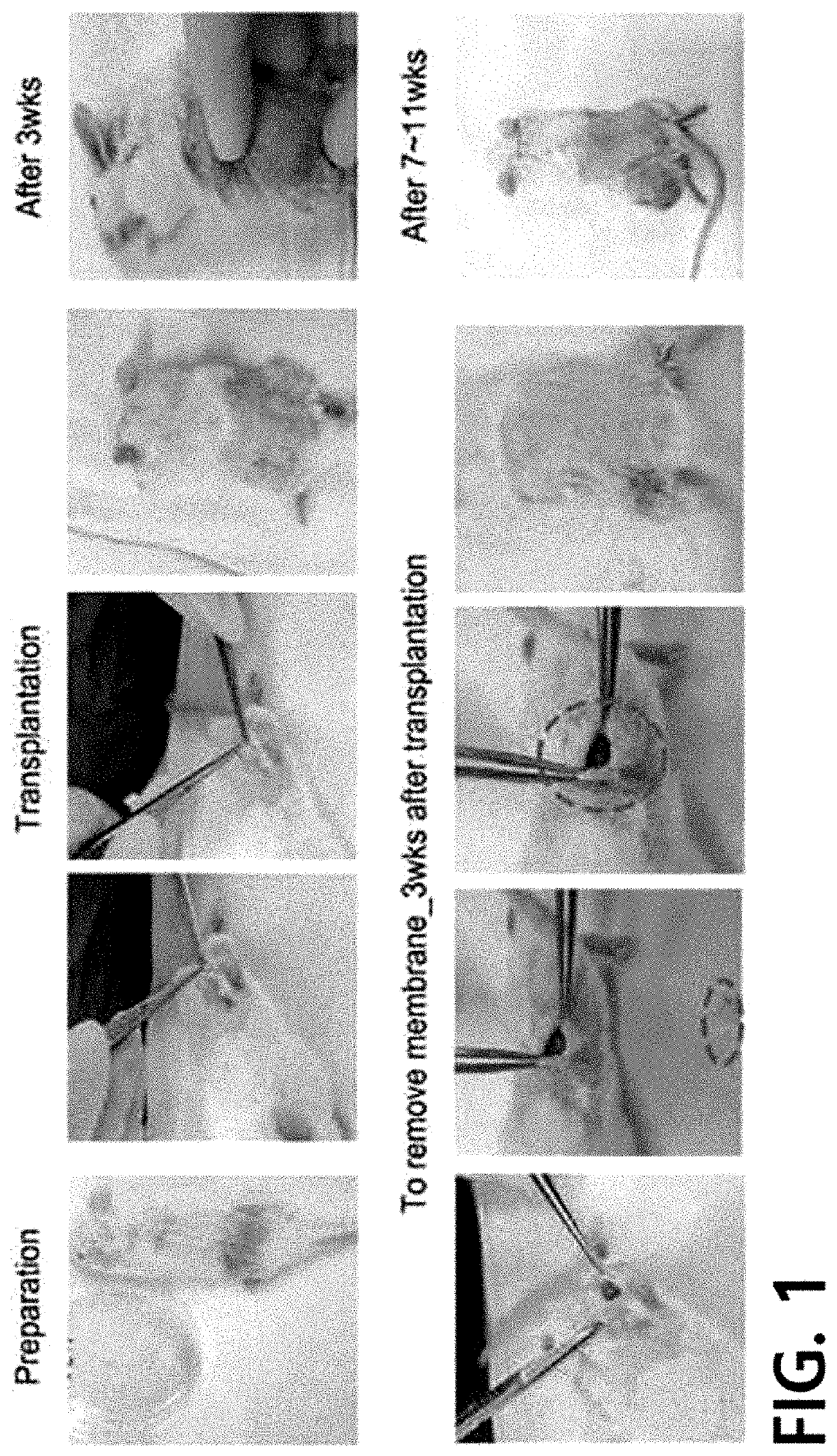
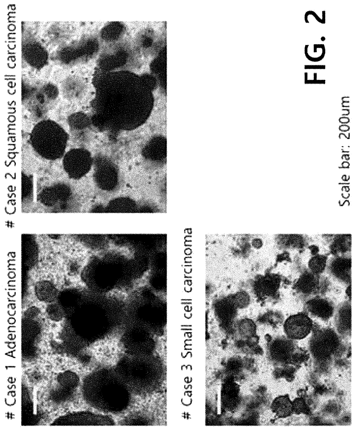
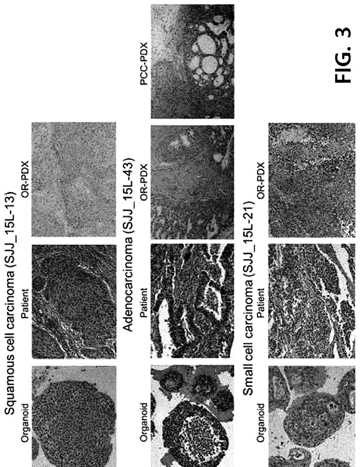
Example 1. 3-Dimensional Culture of Patient-Derived Lung Cancer Cells

[0069]1-1) Cellization of Patient-Derived Lung Cancer Tissue

[0070]The samples in the Preparation Example were cellized according to a known protocol. Specifically, each of the samples in the Preparation Example was placed on a petri dish, and trimming was done using a surgical knife to provide only a cancer tissue portion. The cancer tissue was cut to 1 to 2 mm, transferred to a 15-ml conical tube, and pipetted vigorously 3 to 4 times with a washing solution (DMEM media+0.1% BSA). The resultant was left to stand for 1 minute so that the tissue precipitates due to gravity, and then the supernatant was removed. The above washing step was repeated 3 to 4 times until the tissue becomes clean.

[0071]Then, the same amount of digestion solution as the tissue was added thereto and reaction was allowed to proceed for 40 to 60 minutes in a 37° C. rotary incubator. After 20 minutes, a microscope (40 to 100 magnification) was u...

example 2

n of Organoid and Enzymatic Dissociation

[0077]2-1) Separation of Organoid

[0078]The medium for lung cancer organoid used for the culture in Example 1-2) was placed in a water bath and warmed up. Then, the medium in each well of the 6-well plate was removed. Subsequently, 500 μl of cold PBS was placed in each well, and the organoid-containing Matrigel was removed from the plate. Subsequently, the Matrigel / PBS mixture was transferred to a 15-ml conical tube, and the organoid was separated.

[0079]2-2) Enzymatic Dissociation

[0080]The organoid separated in Example 2-1) was centrifuged with 250×g for 3 minutes at 4° C., and then the supernatant was removed. The resultant was resuspended with PBS, and then centrifugation was performed again with 250×g for 10 minutes at 4° C. to separate the organoid from the Matrigel layer. The supernatant and the Matrigel were removed. Then, 1,000 to 2,000 μl of Trypsin / EDTA was placed in 3 wells of a 6-well plate, and reaction was allowed to proceed for 10...


PUM

PropertyMeasurementUnit
compositionaaaaaaaaaa
biocompatibleaaaaaaaaaa
heterogeneityaaaaaaaaaa

Abstract

The present invention relates to a method for culturing a 3-dimensional lung cancer organoid and a method for preparing a patient-derived xenograft animal model using the same. More specifically, the present invention relates to a method for culturing a 3-dimensional lung cancer organoid, a lung cancer organoid prepared by the method, a medium composition for culturing the lung cancer organoid, a method for preparing a xenograft animal model using the lung cancer organoid, a patient-derived lung cancer organoid xenograft animal model prepared by the method, and a method for analyzing therapeutic efficacy of an anticancer agent and a method for screening an anticancer agent, using the animal model.

Description

TECHNICAL FIELD[0001]The present invention relates to a method for culturing a 3-dimensional lung cancer organoid and a method for preparing a patient-derived xenograft animal model using the same. More specifically, the present invention relates to a method for culturing a 3-dimensional lung cancer organoid, a lung cancer organoid prepared by the method, a medium composition for culturing the lung cancer organoid, a method for preparing a xenograft animal model using the lung cancer organoid, a patient-derived lung cancer organoid xenograft animal model prepared by the method, and a method for analyzing therapeutic efficacy of an anticancer agent and a method for screening an anticancer agent, using the animal model.BACKGROUND ART[0002]In cancer research, cancer models that well represent the patient's characteristics are important. Cancer models that have been used mainly in vitro until now are cancer cell lines. Cancer cell lines are models prepared by adapting patient's cancer t...

Claims


Application Information

Patent Timeline
Patent Type & Authority Applications(United States)
IPC IPC(8): A61K49/00C12N5/09A01K67/027
CPCA61K49/0008C12N5/0693A01K67/0271C12N2513/00C12N2533/50C12N2500/38A01K2227/105C12N2501/113C12N2501/115C12N2501/727A01K2207/12A01K2267/0331C12N2501/11C12N5/0688C12N2509/00C12N2533/90G01N33/57423G01N33/5088G01N2800/52A01K2267/0393A01K67/027C12N2501/50
Inventor JANG, SE JINKIM, MIN SUHSUH, YOUNG AHMUN, HYE MINOH, JU HEE
Owner UNIV OF ULSAN FOUND FOR IND COOPERATION
Why Eureka
Try Eureka