Check patentability & draft patents in minutes with Patsnap Eureka AI!

Biological object detection

a biological object and detection technology, applied in the field of diagnostic assistance system, can solve the problems that the acm approach cannot identify the boundary of a nucleus, and achieve the effect of improving contour detection accuracy, efficient evaluation of shape compactness metrics, and more accurate detection of boundary boundaries

Active Publication Date: 2021-07-22
KONINKLJIJKE PHILIPS NV
View PDF0 Cites 2 Cited by
  • Summary
  • Abstract
  • Description
  • Claims
  • Application Information

AI Technical Summary

Benefits of technology

The proposed approach in this patent improves the accuracy of detecting the boundary of a nucleus in a digital pathology image by using super-pixels and not depending on the color or texture analysis of the image. Super-pixels are contiguous regions of pixels that are defined using a super-pixel segmentation algorithm. This approach helps to segment parts of the nucleus and recognize it in the image. The application of this approach leads to more accurate detection of the boundary of a biological object having a non-homogeneous color or texture distribution.

Problems solved by technology

ACM approaches could fail to identify the boundary of a nucleus.

Method used


Image

  • Biological object detection
  • Biological object detection
  • Biological object detection

Examples


Embodiment Construction

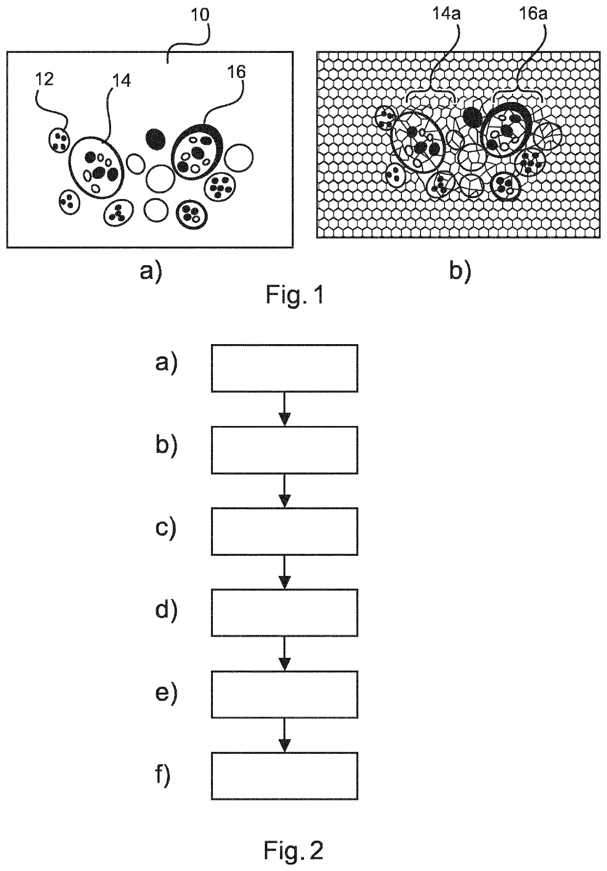
[0054]Nucleus detection is a difficult problem both for H&E, and for immunohistochemistry (IHC) images. A typical approach is to detect initial locations using image intensity, initialize a contour location and shape, and to apply an Active Contour Model (ACM) to define accurately the boundary of cell nuclei. This approach assumes that useful nucleus candidates have already been detected. Additionally, the final boundary of a nucleus from the candidate nuclei can only be found by imposing smoothness on the boundary, as expressed by an energy minimization function.

[0055]This approach may not be the preferred solution in terms of robustness or the algorithm efficiency. Initialization of the contour in the ACM approach can encounter difficulties when the cell under analysis consists of an open structure, in other words, when different parts of the cell or nucleus have a very different colour intensity owing to the ingress of cell cytoplasm, for example.

[0056]In such cases, prior art ap...


PUM


Abstract

As the capabilities of digital histopathology machines grows, there is an increasing need to ease the burden on pathology professionals of finding interesting structures in such images. Digital histopathology images can be at least several Gigabytes in size, and they may contain millions of cell structures of interest. Automated algorithms for finding structures in such images have been proposed, such as the Active Contour Model (ACM). The ACM algorithm can have difficulty detecting regions in images having variable colour or texture distributions. Such regions are often found in images containing cell nuclei, because nuclei do not always have a homogeneous appearance. The present application describes a technique to identify inhomogeneous structures, for example, cell nuclei, in digital histopathology information. It is proposed to search pre-computed super-pixel information using a morphological variable, such as a shape-compactness metric, to identify candidate objects.

Description

FIELD OF THE INVENTION[0001]This invention relates generally to a diagnosis assistance system configured to identify a contour of a biological object in image information, to an image processing method for identifying a contour of a biological object in image information, and additionally a computer program element for controlling a processing unit, and a computer-readable medium.BACKGROUND OF THE INVENTION[0002]Digital pathology seeks to increase the automation of the difficult process of identifying objects in pathology slides containing samples taken from human or animal patients. Digital pathology is difficult because improvements in acquisition hardware have enabled pathology slides of many gigabytes in size to be recorded. Such slides typically contain hundreds of thousands, or millions of targets of potential interest to a medical professional.[0003]One approach to detect the boundary of objects in digital pathology images is via the use of so called “seed regions”. These see...

Claims


Application Information

Patent Timeline
IPC IPC(8): G06T7/00G06T7/11
CPCG06T7/0014G06T2207/30024G06T2207/20036G06T7/11G06T7/0012G06T7/187G06T7/00
Inventor VAREKAMP, CHRISTIAAN
Owner KONINKLJIJKE PHILIPS NV
Features
  • R&D
  • Intellectual Property
  • Life Sciences
  • Materials
  • Tech Scout
  • Unparalleled Data Quality
  • Higher Quality Content
  • 60% Fewer Hallucinations
Learn More