Eureka AIR delivers breakthrough ideas for toughest innovation challenges, trusted by R&D personnel around the world.

Medical imaging data compression and extraction on client side

a technology for medical imaging and data compression, applied in the field of medical imaging data compression and extraction on the client side, can solve the problems of significant delays in transmitting data in at least some instances, prior approaches to handling data can be less than ideal in at least some respects, and the amount of image data has increased, so as to improve the functioning of a computing device, improve the transmission and storage of medical imaging data, and reduce the amount of data

Pending Publication Date: 2021-08-26
ALIGN TECH
View PDF7 Cites 1 Cited by
  • Summary
  • Abstract
  • Description
  • Claims
  • Application Information

AI Technical Summary

Benefits of technology

[0008]The pre-processed 3D volumetric image data may be compressed using any appropriate compression technique. In particular one or more video compression techniques may be used by converting a spatial axis of the 3D volume of the 3D volumetric image data into a time axis to form a time sequence of 2D images and applying a video compression scheme. By compressing the pre-processed 3D volumetric image data, the methods and apparatuses described herein may provide a client-side solution for compressing and / or extracting medical imaging data. The systems and methods described herein may improve the transmission and storing of medical imaging data, by reducing an amount of data to be uploaded to a server.
[0009]In addition, the systems and methods described herein may improve the functioning of a computing device by more efficiently applying a compression scheme and reducing bandwidth by transmitting compressed data. These systems and methods may also improve the field of medical imaging by reducing data storage requirements for computational processes.

Problems solved by technology

However, as medical imaging techniques become more sophisticated, the amount of image data has increased and the prior approaches to handling data can be less than ideal in at least some respects.
Although a dental or orthodontic practitioner may be able to view and manipulate the digital volume to diagnose and treat the region of interest, there can be significant delays in transmitting data in at least some instances.
Although the digital volume may provide an accurate 3D representation, the volumetric data storage and transfer requirements may burdensome and less than ideal in at least some instances.
For example, the CBCT scanner may generate volumetric data, and a large amount of volumetric data may be difficult and time consuming to consistently transfer to a remote location.
Also, it may be more difficult than ideal to store or otherwise archive multiple sets of volumetric data in multiple locations, for example.

Method used


Image

  • Medical imaging data compression and extraction on client side
  • Medical imaging data compression and extraction on client side
  • Medical imaging data compression and extraction on client side

Examples


Embodiment Construction

[0042]The following detailed description and provides a better understanding of the features and advantages of the inventions described in the present disclosure in accordance with the examples disclosed herein. Although the detailed description includes many specific examples, these are provided by way of example only and should not be construed as limiting the scope of the inventions disclosed herein.

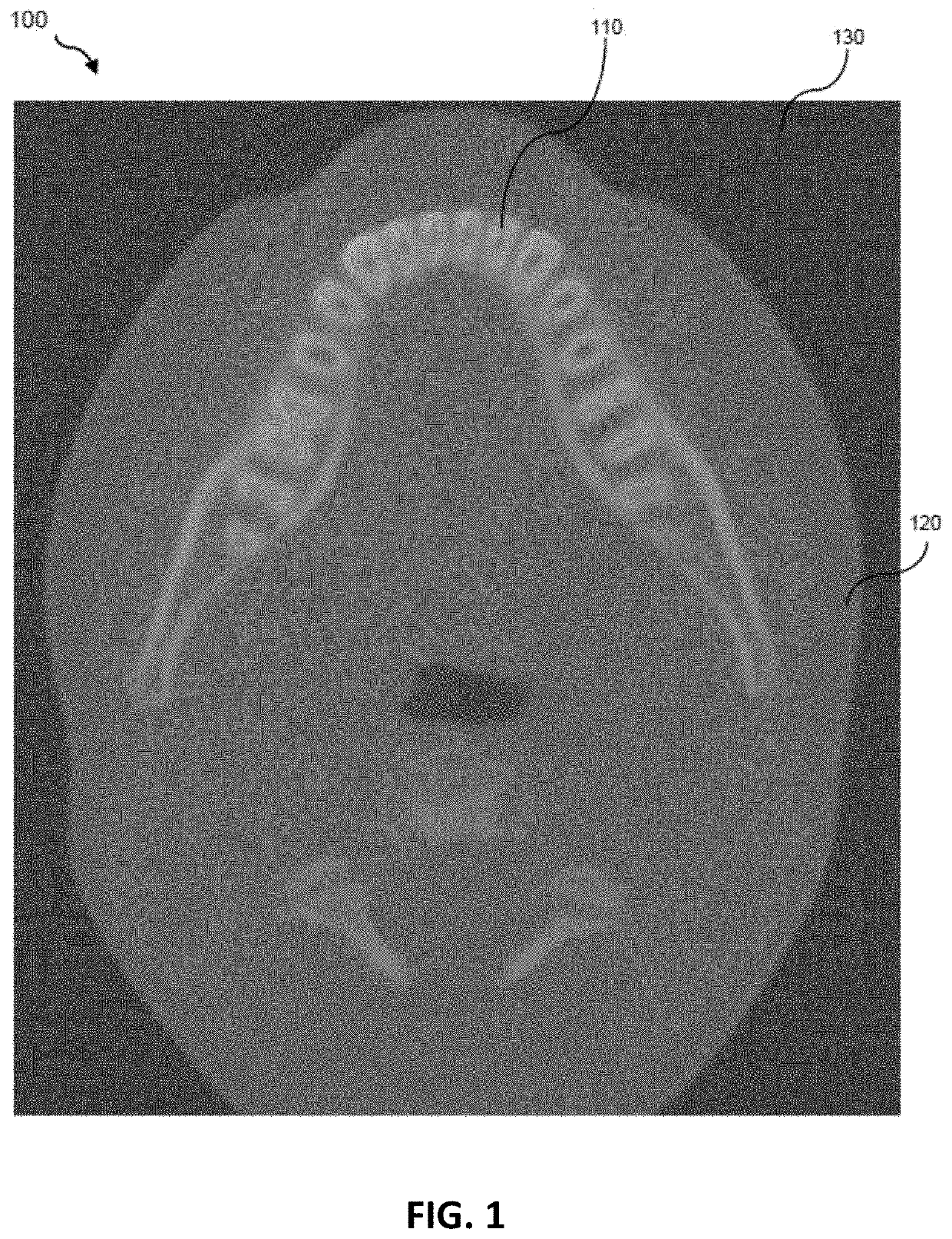
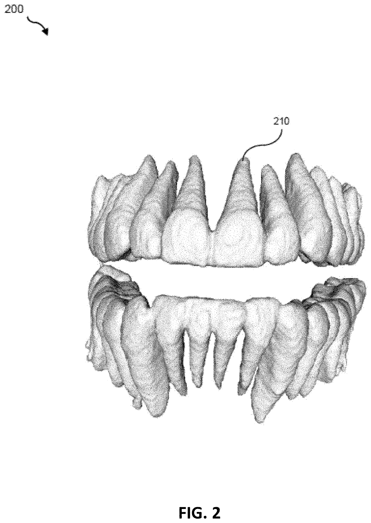
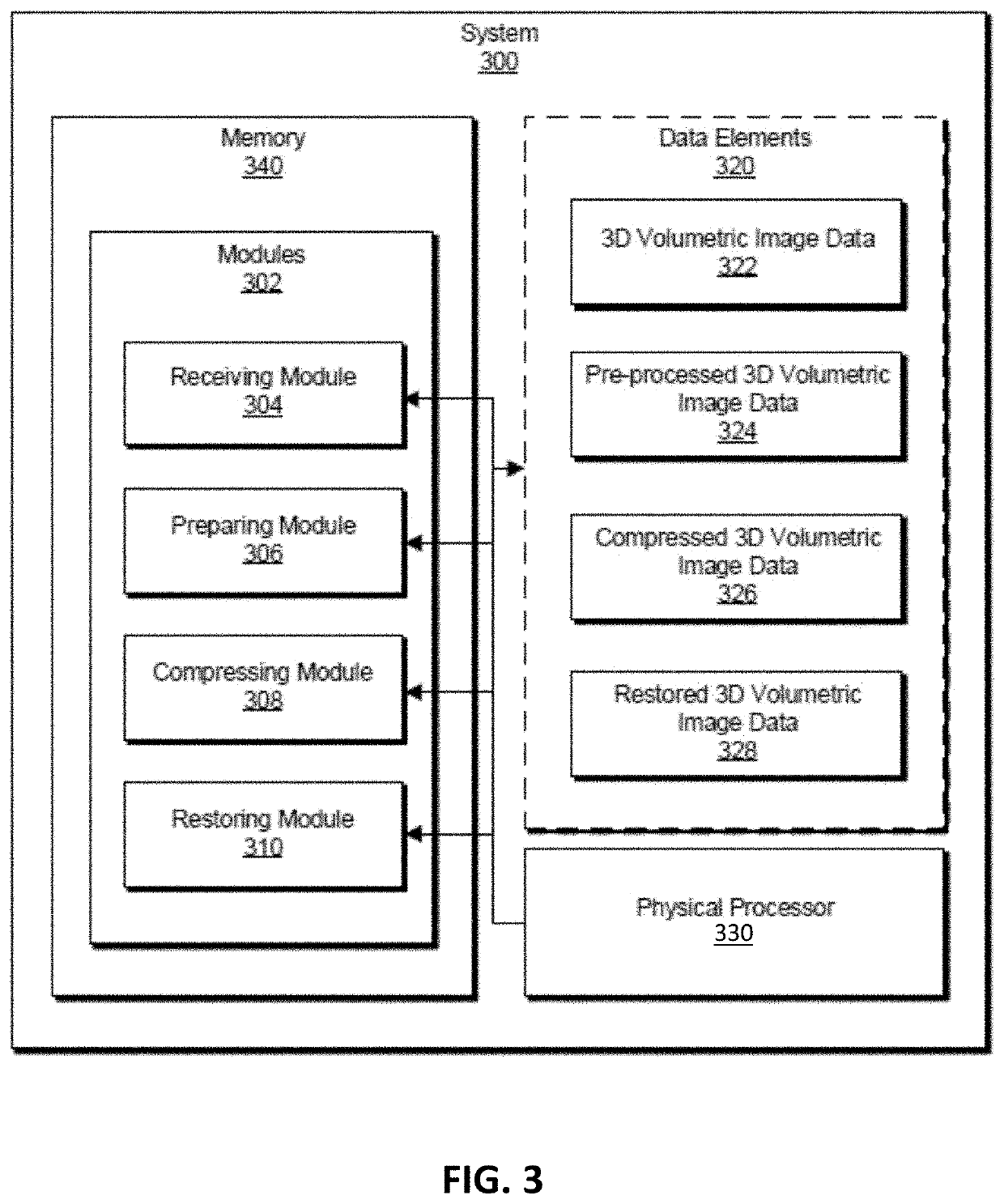
[0043]The following will provide, with reference to FIGS. 1-2, detailed descriptions of imaging and modeling. Theses examples may be specific to medical (e.g., dental) systems and methods, but the methods and apparatuses described herein may be generally applied to any (including non-medical) imaging technique. Detailed descriptions of example systems for compressing and extracting medical imaging data will be provided in connection with FIGS. 3-4. Detailed descriptions of corresponding computer-implemented methods will also be provided in connection with FIG. 5. Detailed descriptions...


PUM


Abstract

Apparatuses and computer-implemented methods for compression and extraction of three-dimensional (3D) volumetric imaging data, including in particular medical and dental imaging data, that may receive the 3D volumetric image data and preparing compressed 3D volumetric image data for compression by analyzing a relevant region of the 3D volumetric image data. The method may further include compressing, into compressed 3D volumetric image data, the pre-processed static 3D volumetric image data using a video compression scheme. Various other methods, systems, and computer-readable media are also disclosed.

Description

CLAIM OF PRIORITY[0001]This patent application claims priority to U.S. Provisional Patent Application No. 62 / 979,325, filed on Feb. 20, 2020, titled “MEDICAL IMAGING DATA COMPRESSION AND EXTRACTION ON CLIENT SIDE,” herein incorporated by reference in its entirety.INCORPORATION BY REFERENCE[0002]All publications and patent applications mentioned in this specification are herein incorporated by reference to the same extent as if each individual publication or patent application was specifically and individually indicated to be incorporated by reference.BACKGROUND[0003]Modern medicine increasingly relies on medical imaging for diagnosing and treating patients. For example, medical imaging may provide visual representations of the interior of a patient's body. Various medical imaging techniques, such as X-ray radiography, CT-scan imaging, magnetic resonance imaging (MRI), and ultrasonography, can be used to produce image data of a patient's body. For example, cone beam computed tomograp...

Claims


Application Information

Patent Timeline
Patent Type & Authority Applications(United States)
IPC IPC(8): G16H30/40G06T5/00G06T5/40G06T7/11H04N19/176H04N19/625H04N19/91G16H30/20A61C9/00
CPCG16H30/40G06T2207/10081G06T5/40G06T7/11H04N19/176H04N19/625H04N19/91G16H30/20A61C9/004G06T2200/04G06T2207/20132G06T2207/10016G06T2207/30036G06T2207/20052G06T5/009H04N19/85G06T5/92
Inventor SLYNKO, YURYCHERNOV, VITALIY VLADIMIROVICHBARYSHNIKOV, ILYA
Owner ALIGN TECH
Why Eureka
Eureka Blog
Learn More