Nucleic acid origami structure encapsulated by capsid units
a nucleic acid and origami technology, applied in the field of viral capsid units encapsulating nucleic acid origami structure, can solve the problems of not being able to efficiently enter the nucleus of particles which do not exhibit high resemblance to viruses, and not being able to efficiently deliver nucleic acid, so as to facilitate nuclear uptake of nucleic acid cargo, efficient and complete nucleic acid delivery
- Summary
- Abstract
- Description
- Claims
- Application Information
AI Technical Summary
Benefits of technology
Problems solved by technology
Method used
Image
Examples
example 1
Assembly of SV40 VP1 on DNA Origami
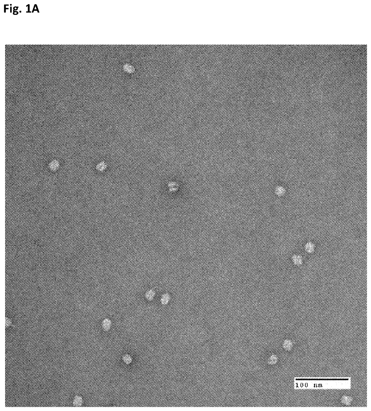
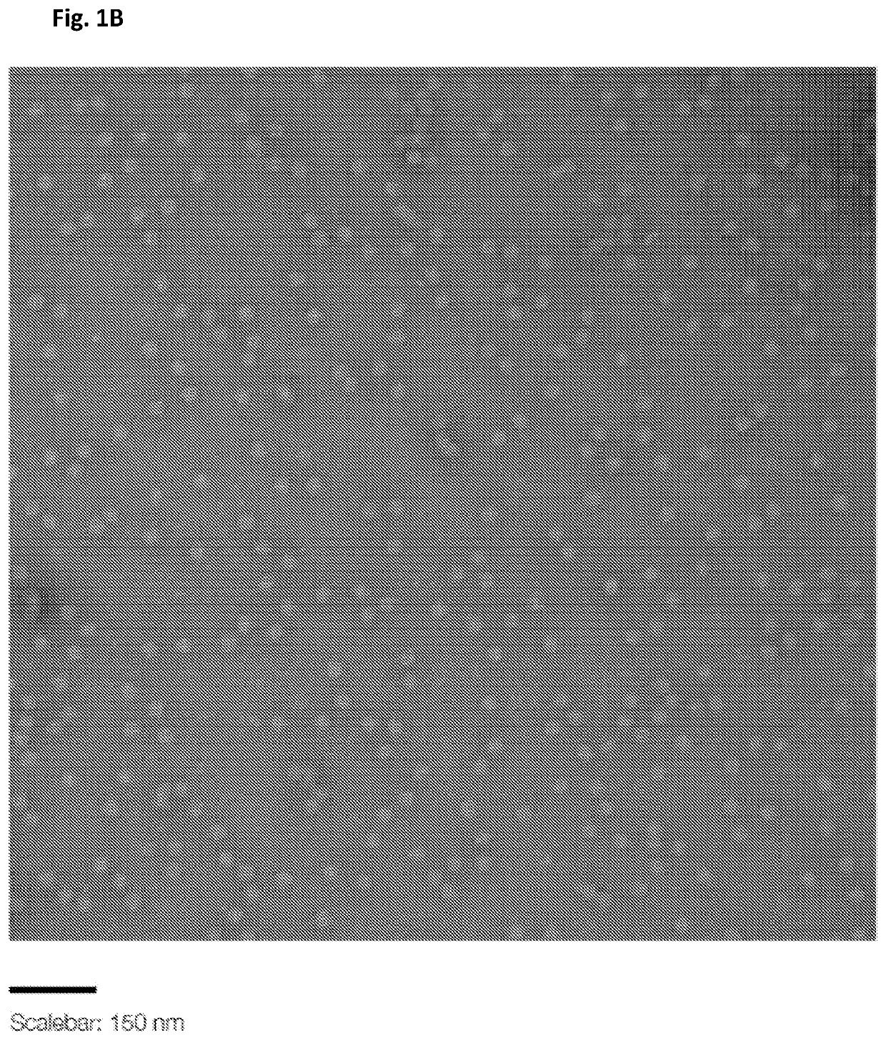
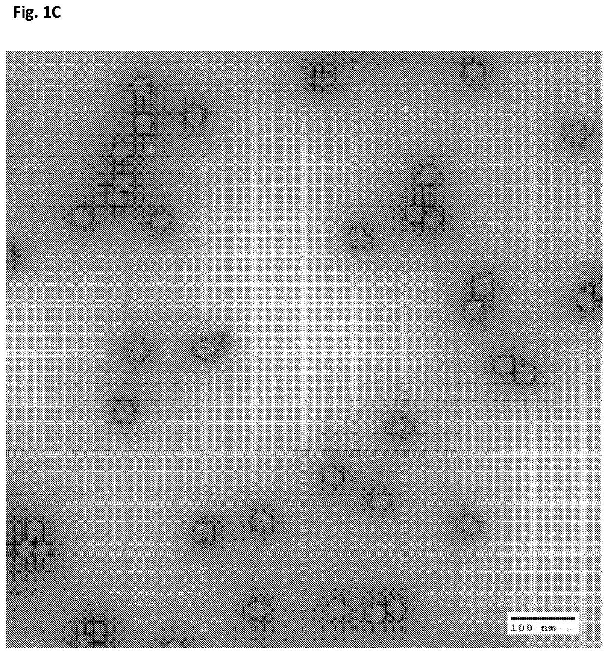
[0113]Origami structure DNA in the form of near-spheres, were used for the particle preparation. Origami DNA structures of the indicated diameters were purchased from tilibit nanosystems, Garching, Germany: 25 nm, 30 nm, 35 nm and 40 nm (shown in the Electron micrographs of FIGS. 1A-D). Additionally, a 30 nm DNA origami structure with 25 staples modified with a Cy5 fluorescent tag was used. All DNA origami structures were incorporated into the particles (VLPs) and used in the herein described experiments.
[0114]Capsid units made of pentamers of SV40 capsid protein VP1 were used for encapsulation of the origami structures. For negative staining TEM, cryo-EM and agarose gel analysis, VLPs were prepared by using 400:1 pentamers / origami structure ratio: (pentamers—4 μM; origami structures—10 nm).
example 2
Formation of Virus-Like Particles
[0115]Negative staining Transmission Electron Microscopy (TEM) was used to confirm the formation of VLPs. VLPs were negatively stained with 2% uranyl acetate and imaged. Assembly of VP1 on 25 nm DNA origami structure leads to formation of 36-38 nm particles (as shown in FIG. 2A). Assembly of VP1 around DNA origami structure of 30 nm diameter leads to formation of 50 nm particles (as shown in FIG. 2B).
[0116]Assembly of VP1 around near-spherical DNA origami structure of 35 nm diameter leads to formation of 50 nm particle as well (As shown in FIG. 2C).
[0117]Assembly of VP1 around near-spherical DNA origami structure of 40 nm in diameter leads to formation of 56 nm particle. (As shown in FIG. 2D).
[0118]The particles obtained by assembly of VP1 around 25 nm and 40 nm DNA origami structures represent entirely new forms of SV40 particles, with non-native capsid sizes, suggesting geometry of T=3 or 4 and T=12, respectively. These results suggest that T, the ...
example 3
Cryo-EM of SV40-Like Particles Assembled on 30 nm Nearly-Spherical DNA Origami Structure
[0119]Cryo-EM 2D micrographs of eight typical particles formed by VP1 assembly on 30 nm near-spherical DNA origami were performed. The resulting particles are practically identical to SV40 in terms of size and shape. Particles are comprised of a shell exhibiting characteristic viral spikes on the circumference (VP1 pentamers) and a core. In some of the cores, the folded strands of the origami structure are visible (seen as stripes), as shown in FIG. 3A. To demonstrate resemblance to native virus, the 3D model of wild type SV40 (PDB entry 1SVA) shows perfect alignment when superimposed on the synthetic particle (FIG. 3B). 2D classification of Cryo-EM micrographs sorts particles into classes of similar particles, each subclass representing an average of several hundred particles. Subtypes are all very similar to each other and exhibiting viral characteristics, as illustrated in FIG. 3C. The 2D clas...
PUM
| Property | Measurement | Unit |
|---|---|---|
| Diameter | aaaaa | aaaaa |
| Diameter | aaaaa | aaaaa |
Abstract
Description
Claims
Application Information
 - R&D
- Intellectual Property
- Life Sciences
- Materials
- Tech Scout
- Unparalleled Data Quality
- Higher Quality Content
- 60% Fewer Hallucinations
Browse by: Latest US Patents, China's latest patents, Technical Efficacy Thesaurus, Application Domain, Technology Topic, Popular Technical Reports.
© 2025 PatSnap. All rights reserved.Legal|Privacy policy|Modern Slavery Act Transparency Statement|Sitemap|About US| Contact US: help@patsnap.com


