
Image processing system and method

a processing system and image technology, applied in the field of image processing system, can solve the problems of reducing the invasive vat, affecting the effect of image enhancement, and unable to operate on pre-operative ct,

Active Publication Date: 2021-10-14
KONINKLJIJKE PHILIPS NV
View PDF11 Cites 0 Cited by
  • Summary
  • Abstract
  • Description
  • Claims
  • Application Information

AI Technical Summary

Benefits of technology

The patent describes a method for improving lung navigation during interventions. It involves detecting specific patterns on the lung surface that are unique to a certain view. These patterns are not significantly changed during lung deflation. By focusing on these patterns, the system provides better contrast and improves the overall visualization of the lung. This makes it easier to navigate the lung and target specific structures, such as lesions. Overall, the system helps to reliably relate pre-operative image information to the intra-operative situation.

Problems solved by technology

One issue with lung surgery, both open thoracotomy and minimally-invasive VATS, is lung deflation.
This makes navigation based, for example, on pre-operative CT difficult.
In VATS, this is no longer possible.

Method used


Image

  • Image processing system and method
  • Image processing system and method
  • Image processing system and method

Examples


Embodiment Construction

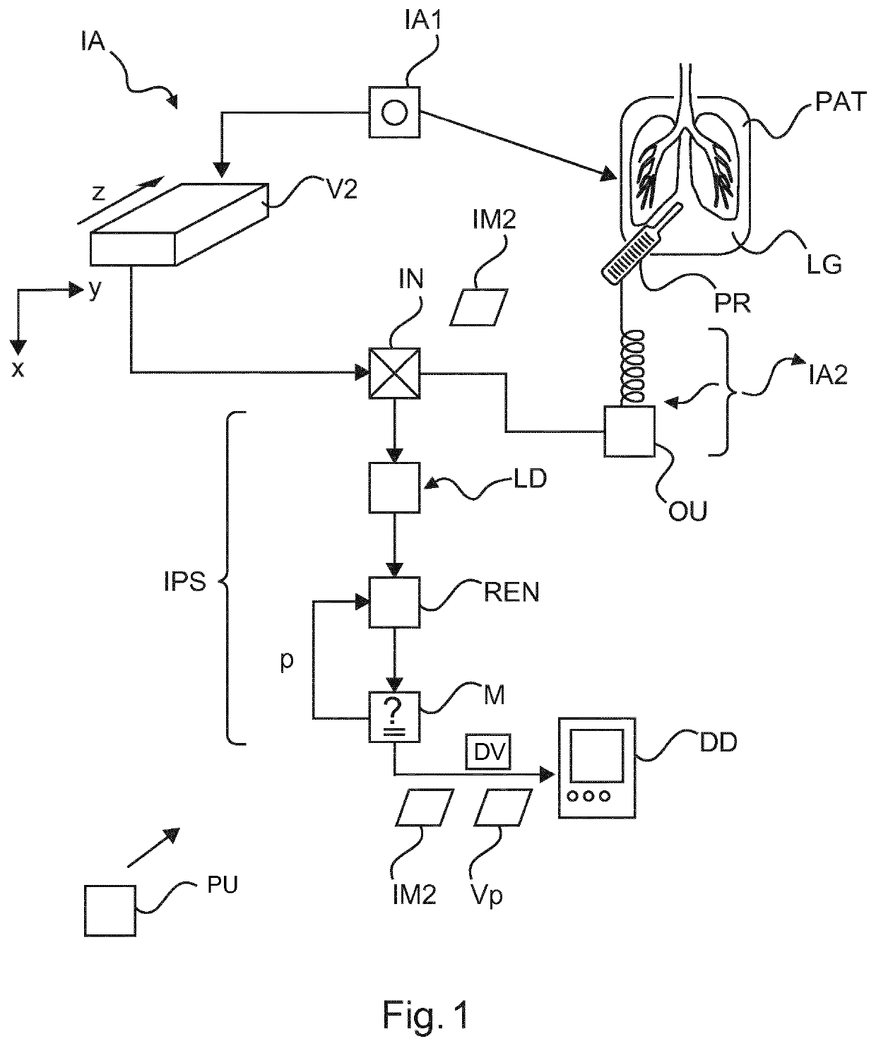
[0047]With reference to FIG. 1, there is shown a schematic block diagram of an imaging arrangement IA as envisaged herein in embodiments.

[0048]The imaging arrangement IA is in particular configured for imaged-based support of lung LG interventions. To this end, the imaging arrangement IA includes two imaging modalities IA1 and IA2, preferably different.

[0049]One of the imaging modalities IA1, also referred to herein as the pre-operative imaging modality IA1, is configured to acquire a preferably volumetric VL image set of a human or animal patient PAT. The volumetric imagery VL includes in particular a representation of the region of interest ROI which includes in particular lung LG. When referring to lung LG herein, this should be construed as a reference to either the left lung, the right lung or to both. Contrast agent may be administered to the patient prior or during imaging with the pre-operative imager IA1.

[0050]The second imaging modality IA2, referred to as the intra-operat...


PUM


Abstract

A System for image processing (IPS), in particular for lung imaging The system (IPS) comprises an interface (IN) for receiving at least a part of a 3D image volume (VL) acquired by an imaging apparatus (IA1) of a lung (LG) of a subject (PAT) by exposing the subject (PAT) to a first interrogating signal. A layer definer (LD) of the system (IPS) is configured to define, in the 3D image volume, a layer object (LO) that includes a representation of a surface (S) of the lung (LG). A renderer (REN) of the system (IPS) is configured to render at least a part of the layer object (LO) in 3D at a rendering view (Vp) for visualization on a display device (DD).

Description

FIELD OF THE INVENTION[0001]The invention relates to an image processing system, to an image processing method, to a computer readable medium and to a computer program element.BACKGROUND OF THE INVENTION[0002]Lung surgery is becoming more and more minimally invasive. For different kinds of intervention, open thoracotomy is no longer necessary. This reduces greatly patient discomfort as open thoracotomy entailed considerable incisions. Instead, in “Video Assisted Thoracoscopic Surgery” (VATS), a type of minimal invasive surgery, an endoscope is introduced through a small incision into the thoracic cavity. The surgeon is then provided with video images transmitted from an endoscope to a monitor, while operating with instruments, which may be introduced through additional, small incisions.[0003]VATS may be used for oncologic procedures, e.g. for removal of a malignant neoplasm (lobectomy, segmentectomy, wedge resection etc.), but can be used also for other reasons, e.g. volume reductio...

Claims


Application Information

Patent Timeline
IPC IPC(8): G06T15/08A61B90/00A61B34/10G06T7/00
CPCG06T15/08A61B90/37A61B34/10G06T2210/41A61B2090/364G06T2207/30061G06T2207/10068G06T7/0012G06T7/32G06T7/33G06T2207/10072G06T2207/10136
Inventor SABCZYNSKI, JÖRGMUELLER, MANFREDWIEMKER, RAFAEL
Owner KONINKLJIJKE PHILIPS NV
Learn More