
Medical image processing apparatus, processor device, endoscope system, medical image processing method, and program

a medical image processing and processor technology, applied in the field of screen display, can solve the problems of difficult to utilize diagnosis assistance that uses the emphasis processing, hinder the observation of an observation target performed by a doctor,

Pending Publication Date: 2021-11-04
FUJIFILM CORP
View PDF0 Cites 3 Cited by
  • Summary
  • Abstract
  • Description
  • Claims
  • Application Information

AI Technical Summary

Benefits of technology

The patent text describes a system that can make a lesion portion easier to see by using an emphasis display. However, this display may interfere with the observation of the lesion portion if the display style is not appropriate. The present invention helps prevent this problem by allowing the user to utilize the emphasis display or classification when observing the observation target, without having it hindle the observation.

Problems solved by technology

However, in the case of detecting a region of interest in a medical image such as an endoscopic image, displaying the region of interest in a screen, classifying the region of interest, and displaying the classification in the screen, notifications of the region of interest and the classification may hinder observation of an observation target performed by a doctor or the like.
However, if the emphasis processing is set in the off state, it becomes difficult to utilize diagnosis assist that uses the emphasis processing.

Method used


Image

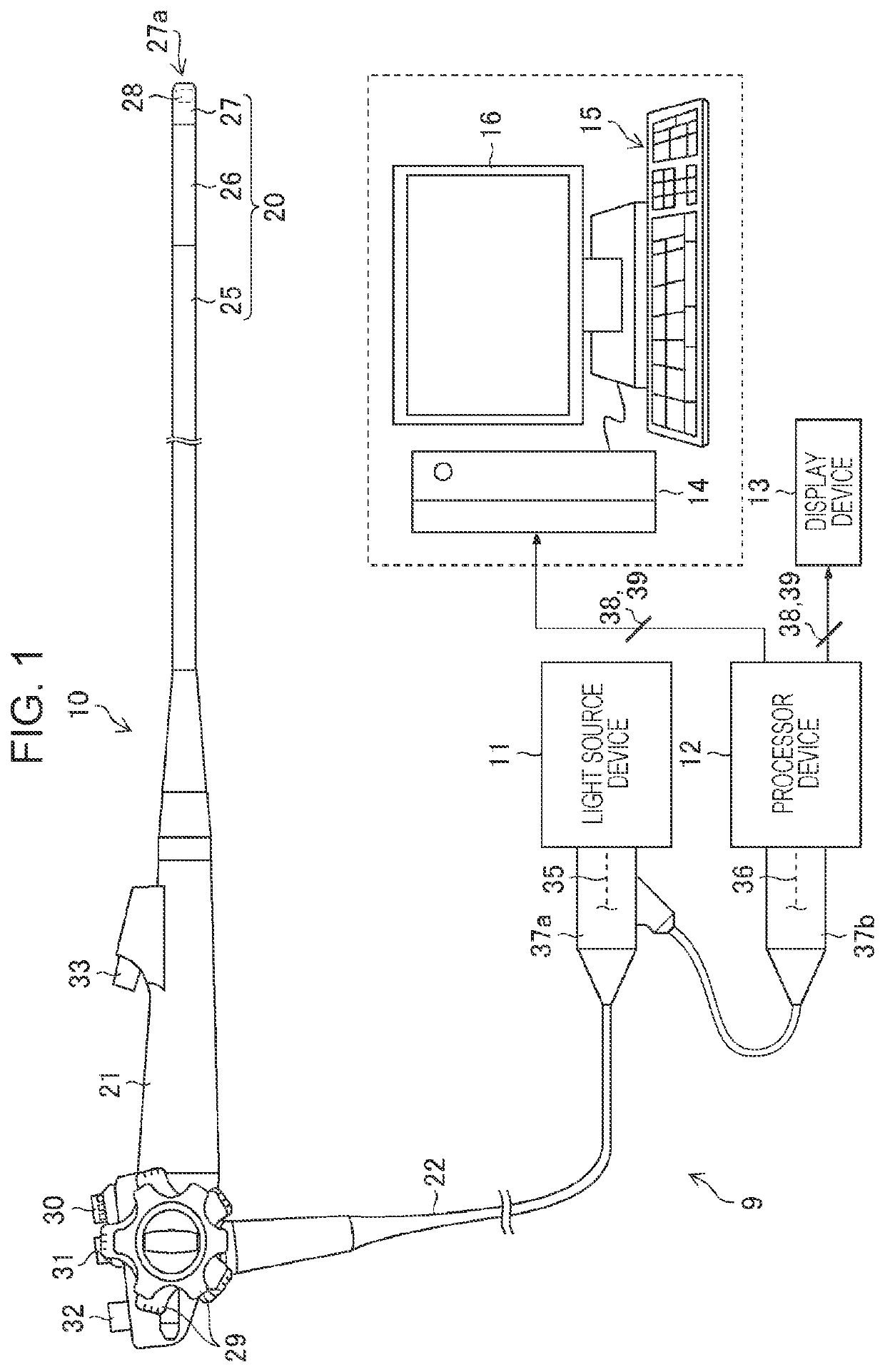
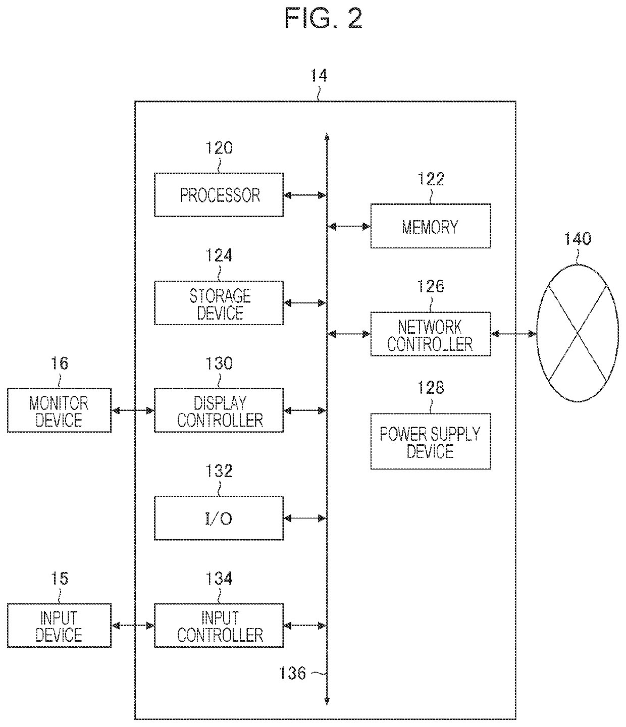
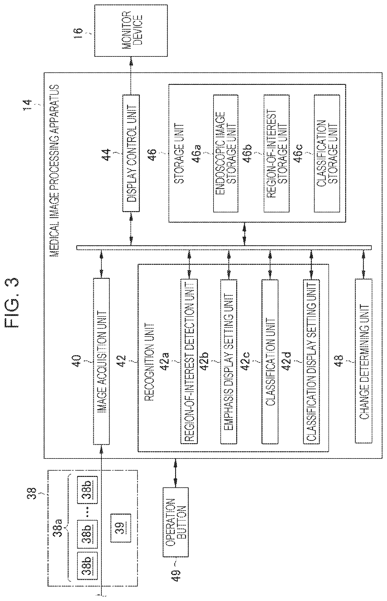
  • Medical image processing apparatus, processor device, endoscope system, medical image processing method, and program
  • Medical image processing apparatus, processor device, endoscope system, medical image processing method, and program
  • Medical image processing apparatus, processor device, endoscope system, medical image processing method, and program

Examples


first modification

[0237]A first example of the specific wavelength range is a blue range or a green range in a visible range. The wavelength range of the first example includes a wavelength range of 390 nm or more and 450 nm or less or a wavelength range of 530 nm or more and 550 nm or less, and the light of the first example has a peak wavelength in the wavelength range of 390 nm or more and 450 nm or less or the wavelength range of 530 nm or more and 550 nm or less.

second modification

[0238]A second example of the specific wavelength range is a red range in the visible range. The wavelength range of the second example includes a wavelength range of 585 nm or more and 615 nm or less or a wavelength range of 610 nm or more and 730 nm or less, and the light of the second example has a peak wavelength in the wavelength range of 585 nm or more and 615 nm or less or the wavelength range of 610 nm or more and 730 nm or less.

third modification

[0239]A third example of the specific wavelength range includes a wavelength range in which an absorption coefficient is different between oxyhemoglobin and deoxyhemoglobin, and the light of the third example has a peak wavelength in the wavelength range in which the absorption coefficient is different between oxyhemoglobin and deoxyhemoglobin. The wavelength range of this third example includes a wavelength range of 400±10 nm, a wavelength range of 440±10 nm, a wavelength range of 470±10 nm, or a wavelength range of 600 nm or more and 750 nm or less, and the light of the third example has a peak wavelength in the wavelength range of 400±10 nm, the wavelength range of 440±10 nm, the wavelength range of 470±10 nm, or the wavelength range of 600 nm or more and 750 nm or less.


PUM


Abstract

A medical image processing apparatus includes a recognition unit that detects a region of interest from a medical image and classifies the region of interest, a display control unit that causes emphasis display of the region of interest and a classification for the region of interest to be displayed in a screen for displaying the observation target, and a change determining unit that determines at least any of whether to change a display style of the emphasis display or whether to change a display style of the classification. The display control unit changes the display style of the emphasis display in the case where it is determined to change the display style of the emphasis display and changes the display style of the classification in the case where it is determined to change the display style of the classification.

Description

CROSS-REFERENCE TO RELATED APPLICATIONS[0001]This application is a Continuation of PCT International Application No. PCT / JP2019 / 041947 filed on Oct. 25, 2019, which claims priority under 35 U.S.C § 119(a) to Japanese Patent Application No. 2019-032944 filed on Feb. 26, 2019. Each of the above application(s) is hereby expressly incorporated by reference, in its entirety, into the present application.BACKGROUND OF THE INVENTIONField of the Invention[0002]The present invention relates to a medical image processing apparatus, a processor device, an endoscope system, a medical image processing method, and a program, and more particularly to screen display.Description of the Related Art[0003]A common endoscope system radiates illumination light from a tip of an insertion section of an endoscope, images an observation target by using an imaging element, and obtains image information of the observation target. An image obtained by imaging is displayed on a monitor. A user such as a doctor o...

Claims


Application Information

Patent Timeline
Patent Type & Authority Applications(United States)
IPC IPC(8): G06K9/00G06K9/32G06K9/62G06T3/40G06T11/60G06F3/14G06T7/00A61B1/00G06V10/25
CPCG06K9/00671G06K9/3233G06K9/6267G06K9/6232G06T3/40G06T2207/30004G06F3/14G06T7/0012A61B1/00045G06K2209/05G06T2207/10068G06T11/60G02B23/24G09G2380/08G09G2340/12G06T11/00A61B1/0005A61B1/000094G06V10/25G06V10/82G06V2201/031G06V20/20G06V2201/03G06F18/24
Inventor OOSAKE, MASAAKIKAMON, SHUMPEI
Owner FUJIFILM CORP
Learn More