
Photoselective non-invasive targeted genomic and epigenomic sequencing of spatially-defined cells or subcellular regions

a targeted, genomic technology, applied in the direction of hydrolases, microorganism testing/measurement, biochemistry apparatus and processes, etc., can solve the problems of inability to achieve subcellular resolution, insensitive to morphological changes, lack of tractability of dispersed target cells,

Pending Publication Date: 2022-07-28
THE BROAD INST INC +1
View PDF2 Cites 0 Cited by
  • Summary
  • Abstract
  • Description
  • Claims
  • Application Information

AI Technical Summary

Benefits of technology

The patent text describes a method for non-invasive sequencing of genomic and epigenomic DNA from tissue samples using light-activated probes. The method involves fixing the tissue sample with a fixing agent, staining it with a fluorescent dye, and preparing a library of DNA fragments. The DNA fragments are then targeted with a near-UV light to unblock the adapters in a specific region of interest. The unblocked adapters are purified and amplified for sequencing. The method can be used for various applications such as diagnostics, therapeutics, and research. The permeability of the tissue sample can be increased using a permeabilizing agent before preparing the library. The DNA fragments can be selected from various DNA molecules such as genomic DNA, mitochondrial DNA, viral DNA, and bacterial DNA. The method is compatible with established Tn5-transposase based library preparations and can generate an image of the tissue sample.

Problems solved by technology

While advancements in methods to effectively profile genomic and epigenomic features have increasingly provided deeper understanding of cell development and disease, such methods, including established DNA sequencing library preparations, usually require cellular dissociation, and are insensitive to morphological features such as the arrangement of cells within a tissue or subcellular structures.
Previous methods for collecting DNA from specific spatial regions, such as laser capture and CHEX-seq (chromatin exposed sequencing), have confronted challenges such as a contamination from neighboring cells during targeted cell excision, inability to achieve subcellular resolutions, lack of tractability for dispersed target cells, and a lack of versatility to measure multiple features from the sample.

Method used


Image

  • Photoselective non-invasive targeted genomic and epigenomic sequencing of spatially-defined cells or subcellular regions
  • Photoselective non-invasive targeted genomic and epigenomic sequencing of spatially-defined cells or subcellular regions
  • Photoselective non-invasive targeted genomic and epigenomic sequencing of spatially-defined cells or subcellular regions

Examples


example 1

ctive Sequencing Provides Spatially Targeted Sequence Data

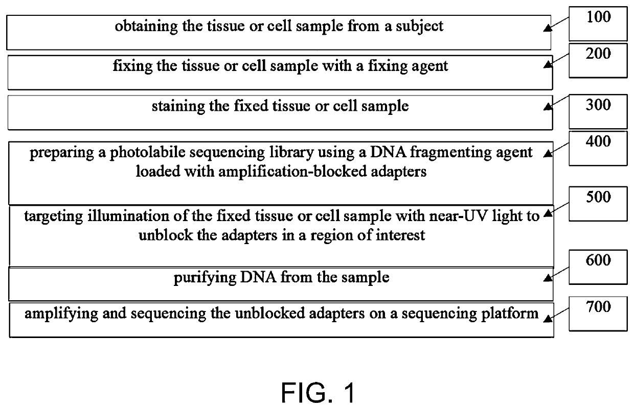
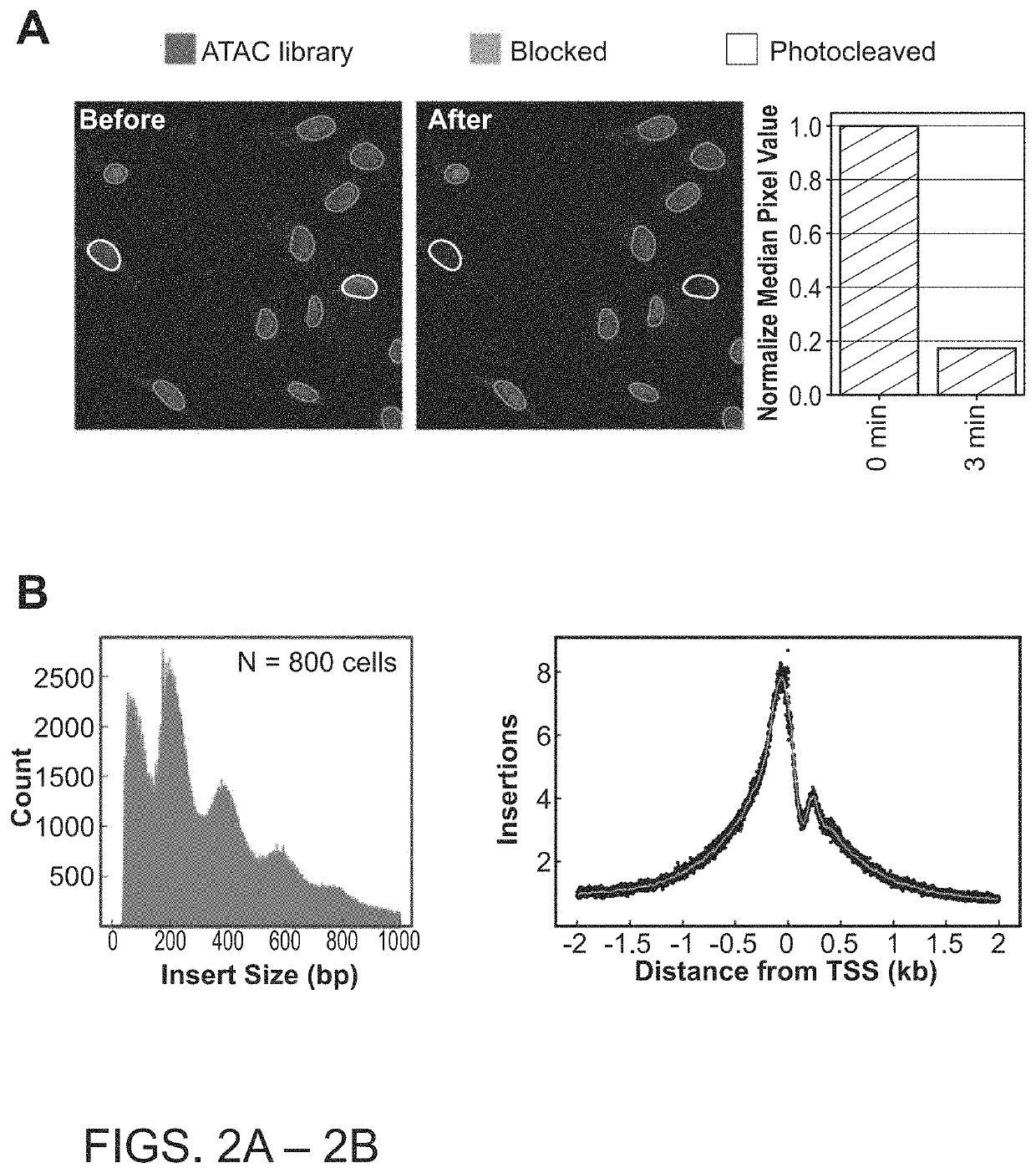
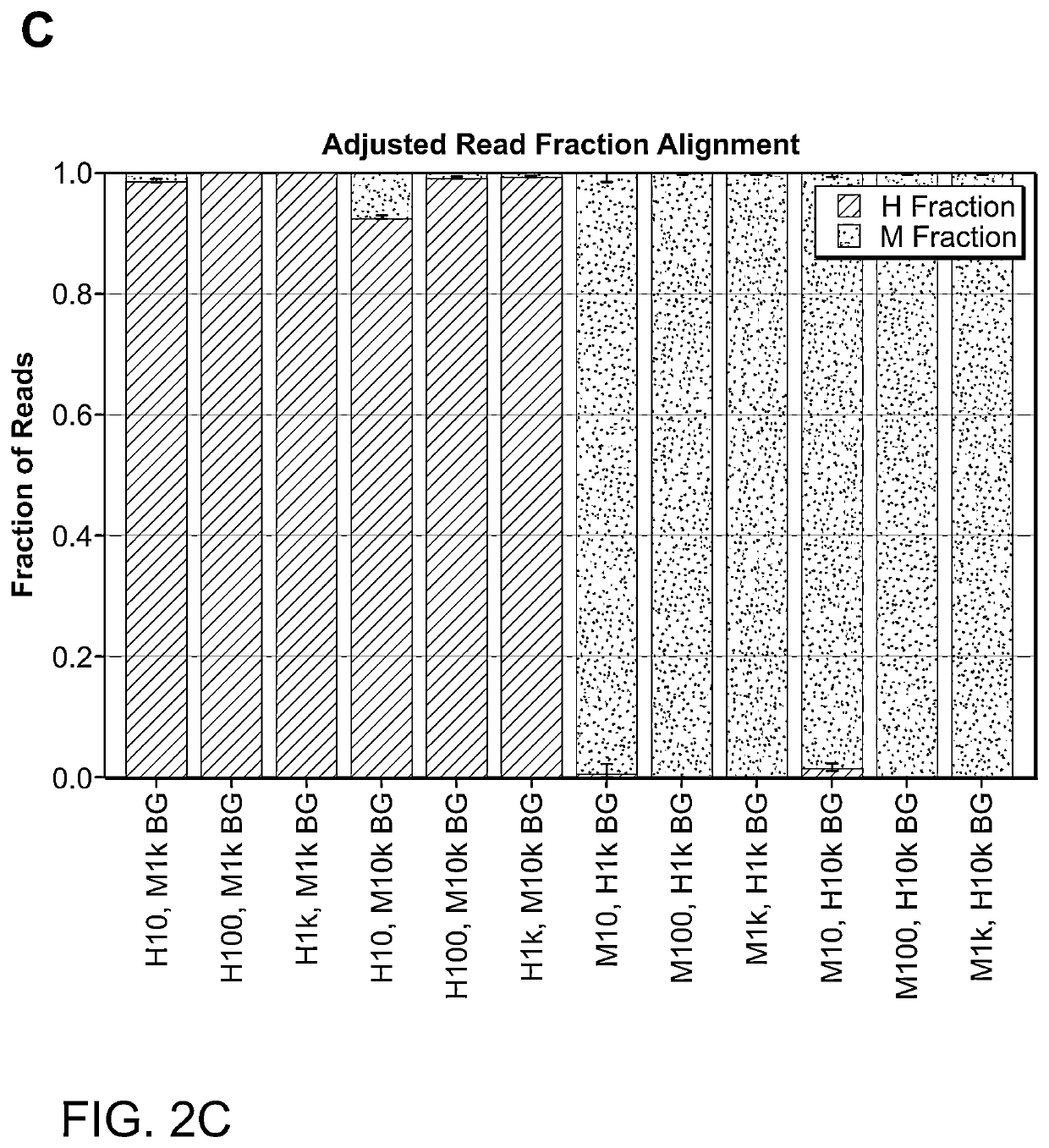
[0163]The techniques herein provide photoselective sequencing, a sequencing methodology that enables non-invasive targeted genomic and epigenomic sequencing of spatially-defined cellular or subcellular regions of tissues or cell populations or cells using light-activated probes. In particular, the disclosure provides in-situ tagmentation with blocked adapters consisting of an oligonucleotide sequence conjugated to a fluorophore via a photocleavable spacer. Photo-induced cleavage of the spacer removes the fluorophore and uncages a 5′ phosphate group that enables ligation of secondary adapters, which are subsequently used to amplify the selected DNA fragments after digestion of the sample. Advantageously, photoselective sequencing provides diffraction-limited resolution and straightforward compatibility with varied genomic and epigenetic sequencing libraries. Additionally, photoselective sequencing is not limited to spatially l...

example 2

and Methods

Sample Protocol for Photoselective Sequencing in Cells and Tissues:

[0168]Unless otherwise specified all steps are carried out at room temperature in a volume of solution that is large enough to completely cover the sample.

Sample Preparation:

[0169]Adherent cells are cultured in an appropriate growth medium and split onto an imaging-compatible dish or slide. For tissues, 10 uM sections of a fresh-frozen sample are collected and placed onto a glass-bottom imaging dish or microscope slide. The tissue is lightly melted onto the glass by touching the underlying glass briefly with one finger.

Fixation and Permeabilization (Cells):

[0170]Cells are rinsed once in 1×PBS, then lightly fixed for 10 minutes at room temperature in 0.4% paraformaldehyde (PFA). Excess PFA is removed from the sample by washing 3 times for 5 minutes each with 1×PBS. Next, cells are permeabilized by incubating in 0.5% Triton X-100 in 1×PBS for 10 minutes at room temperature. The sample is again washed three t...


PUM

PropertyMeasurementUnit
diameteraaaaaaaaaa
temperatureaaaaaaaaaa
thicknessaaaaaaaaaa

Abstract

The present disclosure relates to methods aimed towards non-invasive targeted genomic and epigenomic sequencing of spatially-defined cellular or subcellular region. More particularly, the present disclosure relates to methods of using photoselection to achieve non-invasive targeted genomic and epigenomic sequencing of spatially-defined cellular or subcellular regions, via the use of light-activated probes.

Description

CROSS-REFERENCE TO RELATED APPLICATION[0001]The present application is related to and claims priority under 35 U.S.C. § 119(e) to U.S. provisional patent application No. 63 / 141,342, entitled “Photoselective Non-Invasive, Targeted Genomic and Epigenomic Sequencing of Spatially-Defined Cells or Subcellular Regions,” filed Jan. 25, 2021. The entire content of the aforementioned patent application is incorporated herein by this reference.FIELD OF THE DISCLOSURE[0002]The present disclosure relates to methods aimed towards non-invasive targeted genomic and epigenomic sequencing of spatially-defined cellular or subcellular regions. More particularly, the present disclosure relates to methods of using photoselection for non-invasive targeted genomic and epigenomic sequencing of spatially-defined cellular or subcellular regions using light-activated probes.SEQUENCE LISTING[0003]The instant application contains a Sequence Listing which has been filed electronically in ASCII format and is here...

Claims


Application Information

Patent Timeline
Patent Type & Authority Applications(United States)
IPC IPC(8): C12Q1/686G01N1/30C12N9/22
CPCC12Q1/686C12N9/22G01N1/30C12Q1/6806C12N15/10C12Q2521/507C12Q2523/319C12Q2525/191
Inventor CHEN, FEIMANGIAMELI, SARAHCHEN, HAIQI
Owner THE BROAD INST INC
Learn More