Donor overlay for treatment or alleviation of anterior corneal disorders

a corneal disorder and donor technology, applied in the field of ophthalmology, can solve the problems of corneal perforation, increased risk of postoperative complications, and complex surgical techniqu

Pending Publication Date: 2022-09-22
NIIOS USA INC
View PDF0 Cites 0 Cited by
  • Summary
  • Abstract
  • Description
  • Claims
  • Application Information

AI Technical Summary

Benefits of technology

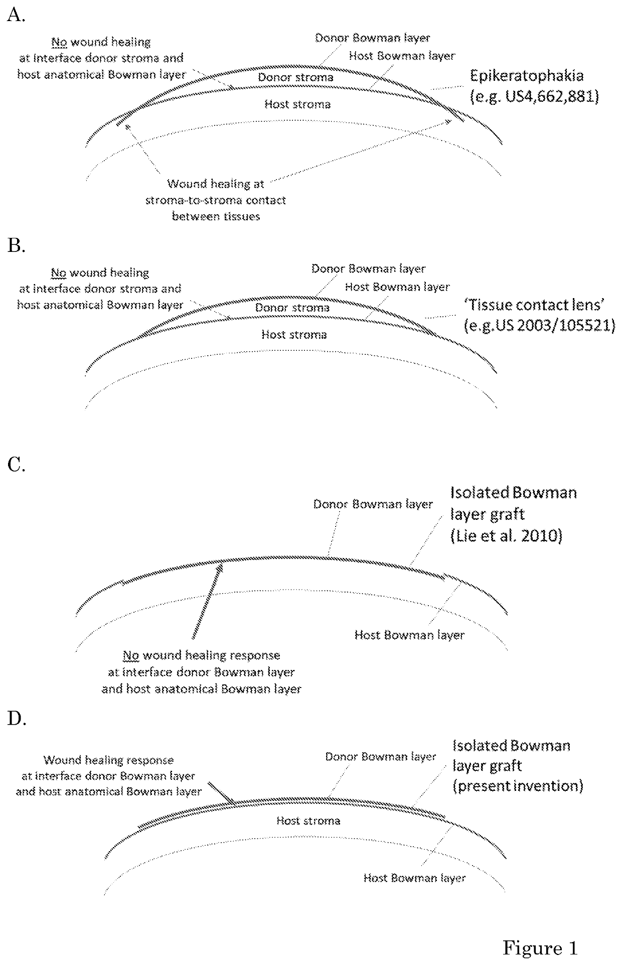
[0047]To overcome the lack of a sufficient wound healing response, the inventor now shows the use of an isolated donor Bowman layer (i.e. a Bowman layer graft devoid of donor stroma) positioned onto the patient's anatomical Bowman layer, since it was hypothesized that it was the donor stroma that interfered with or blocked the wound healing response between a Bowman layer and stromal tissues. In other words, if an (isolated) Bowman layer graft is positioned onto the patient's own Bowman layer (FIG. 1D and FIG. 4), a different type of wound healing response is elicited by a mechanism that is hypothesized not to involve keratocytes, but inflammatory cells derived from the tear film. The wound healing induces tissue contraction and fixation, but also remodeling of the central cornea resulting in improved central corneal clarity.
[0086]A method according to the invention can be performed as follows. From the host cornea, corneal epithelium is carefully removed to denude the recipient BL or in absence thereof, the anterior corneal stroma. The epithelium can for instance be removed by spatula abrasion under topical anesthesia. Pre-dissected donor tissue (preferably BL) at any diameter and either directly after harvesting or after having been stored by any of the methods mentioned above, are removed from the sterile vial, container and / or seal. The donor tissue is then first rehydrated, for example with BSS, and preferably stained with a vital dye as described herein above. The assembly of an multi-layered overlay is preferably performed in an eye bank, for instance. as described herein above, but in the case of a multi-layered overlay or composition according to the invention, the different layers of donor tissue can also be combined during or as part of the surgical procedure itself, preferably after rehydration and either before or after staining with a vital dye. An advantage of a surgical method of the invention is that it is not necessary to suture the overlay or composition onto or into tissue of the eye of the recipient. Hence, in a preferred embodiment, the overlay or composition is positioned sutureless on the corneal tissue of the recipient subject. The overlay or composition is thus positioned onto the cornea of the recipient subject and preferably allowed to ‘dry in’. Positioning the overlay or composition is for instance done using surgical forceps or a spatula. A soft bandage lens is then preferably positioned onto the eye.

Problems solved by technology

However, virtually all treatment modalities known today involve sutured fixation of such tissues which in turn is associated with secondary inflammation and all associated risks thereof.
Most techniques, including the so-called ‘sutureless’ techniques are further invasive which may introduce further risk of postoperative complications.
Although such procedure is associated with less intraoperative and postoperative complications as compared to previously used techniques, the procedure remains of an invasive nature involving the creation of a mid-stromal pocket in the recipient cornea.
In addition, the technique is surgically complex and prone to corneal perforation during surgery.

Method used


Image

  • Donor overlay for treatment or alleviation of anterior corneal disorders
  • Donor overlay for treatment or alleviation of anterior corneal disorders
  • Donor overlay for treatment or alleviation of anterior corneal disorders

Examples


case 3

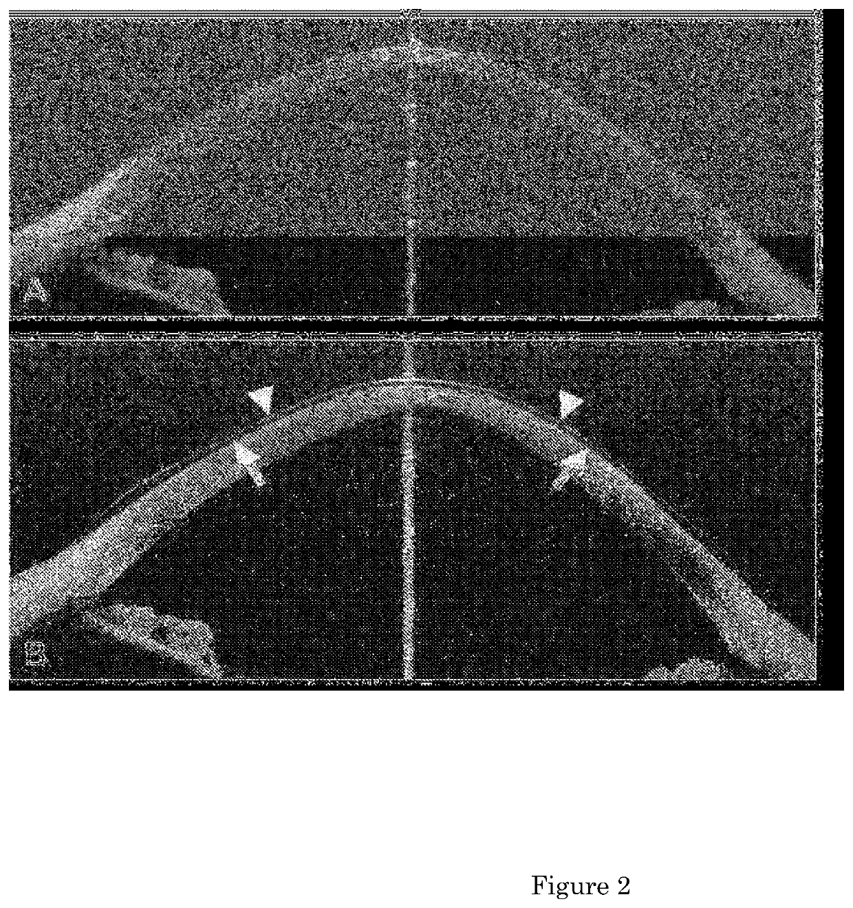
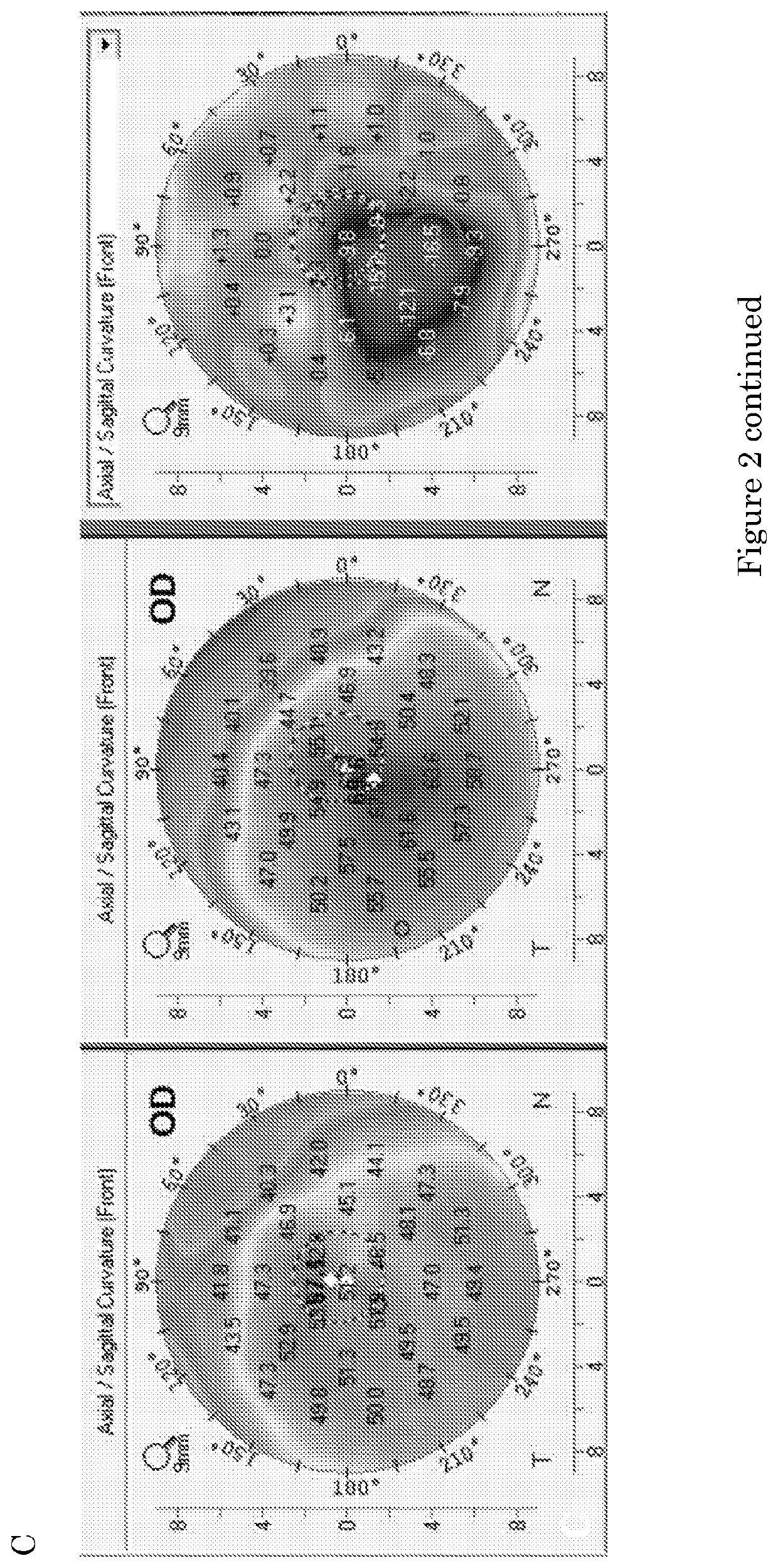
with moderate keratoconus (about 55 diopters) had a 9.0 mm multi-layer BL overlay positioned onto the host remnant BL under topical anesthesia. Six months later, the cornea showed up to 11 diopters of flattening of the anterior corneal surface and thereafter, topography images appeared stable over time.

case 4

with post-excimer laser (LASIK) ectasia (about 50 diopters) had a 9.0 mm single BL overlay positioned onto the host BL under topical anesthesia. Three months later, the cornea showed up to 5 diopters of flattening of the anterior corneal surface and thereafter, topography images appeared stable over time.

case 5

with wound dehiscence >15 years after penetrating keratoplasty had a 9.0 mm single BL overlay positioned onto the central graft under local anesthesia. At six months, topography imaging showed a more regular anterior corneal surface (K-value re-distribution).


PUM

PropertyMeasurementUnit
diameteraaaaaaaaaa
diameteraaaaaaaaaa
thicknessaaaaaaaaaa

Abstract

The invention relates to methods for the treatment or alleviation of an anterior corneal disorder in a subject in need thereof comprising removing corneal epithelial cells from an eye of said subject without removing any corneal tissue or other ocular tissue located posterior to the corneal epithelial cell layer; and positioning an overlay comprising a Bowman layer (BL), Descemet membrane (DM) and / or crystalline lens capsule on the anterior surface of said corneal tissue located posterior to the corneal epithelial cell layer. The invention further relates to freeze-dried and / or gamma irradiated Bowman layer, Descemet membrane and / or crystalline lens capsule and compositions comprising the same that are useful in such methods.

Description

FIELD OF THE INVENTION[0001]The invention relates to the field of ophthalmology, in particular to compositions and tissues for use in eye surgery, especially for treatment or alleviation of anterior corneal disorders.BACKGROUND OF THE INVENTION[0002]Anterior corneal disorders including corneal curvature disorders and / or corneal contour changes, corneal surface disease, corneal defects, refractive errors of the eye benefit from a surgical procedure that allows for transplantation or implantation of donor corneal tissue, to (partially) restore the corneal anatomy, stability and optical quality, and therefore the visual function of the eye. However, virtually all treatment modalities known today involve sutured fixation of such tissues which in turn is associated with secondary inflammation and all associated risks thereof. Most techniques, including the so-called ‘sutureless’ techniques are further invasive which may introduce further risk of postoperative complications.[0003]Traditio...

Claims


Application Information

Patent Timeline
Patent Type & Authority Applications(United States)
IPC IPC(8): A61F2/14A61L27/36
CPCA61F2/1451A61L27/3604A61L27/3666A61L27/3691A61L2430/16A61F2/142
Inventor MELLES, GERRIT REINOLD JACOB
Owner NIIOS USA INC
Why Eureka
Try Eureka