
Artificial intelligence-based colonoscopic image diagnosis assisting system and method

a colonoscopy and image diagnosis technology, applied in the field of medical image diagnosis assisting apparatus and method using an automated system, can solve the problems of reducing the confidence of clinicians in the clinical usefulness of this segmentation technique, unable to fully accept and adopt by users, and no visible results have yet been revealed

Pending Publication Date: 2022-09-22
INFINITT HEALTHCARE CO LTD
View PDF0 Cites 1 Cited by
  • Summary
  • Abstract
  • Description
  • Claims
  • Application Information

AI Technical Summary

Benefits of technology

The present invention provides evaluation scores for various medical image diagnosis algorithms, including confidence and accuracy scores, to improve the accuracy of medical image diagnosis results. Additionally, a method is provided to detect various lesions in real time via artificial intelligence, which can help other examiners check polyp candidates again later by providing the locations of the polyps in a colonoscopic / colonoscopy path. This technology helps to improve the detection rate of polyps in the colon and ultimately enhance the accuracy of colonoscopy examinations.

Problems solved by technology

However, on the other hand, the insides of recent artificial neural networks, such as deep learning and a CNN, approximate black boxes, and thus there is reluctance for a user to fully accept and adopt them even when acquired results are excellent.
However, no visible results have yet been revealed.
However, descriptive information (explanation) about the criteria for the selective application of the segmentation algorithms cannot be derived from the related art document, and thus a problem arises in that it is difficult to increase a clinician's confidence in the clinical usefulness of this segmentation technique.
However, in Korean Patent No. 10-1938992, an artificial neural network derives feature information by itself, and no verification is made as to whether or not the extracted feature information is clinically useful information.
Accordingly, there is little evidence that humans can recognize the above information as a description of the diagnosis result of artificial neural networks.
A similar problem is still present in a medical image diagnosis process in that it is difficult to have clinical confidence in a process in which an artificial intelligence diagnosis system that operates like a black box generates a result.
During colonoscopy, lesions are often overlooked in the course of examination in a narrow path due to causes such as the resolution of an image, a view in which a user does not intuitively perceive a three-dimensional effect, a blind spot, user error, or the like.
In particular, in group health checkups, doctors often exhibit signs of fatigue due to continuous and repetitive procedures, so that lesions may not be sufficiently detected.
Therefore, human error may be combined with the cause in terms of the characteristics of images and cause the mission of lesions, which may negatively affect the medical results of examinations.
However, in the case of deep learning-based artificial intelligence, the fact that there is a black box that prevents a user from determining whether or not a result provided from an operation accidentally exhibits high performance and whether or not a determination process appropriate for a corresponding task has been performed limits the applicability of the deep learning-based artificial intelligence.
In contrast, the use of rule-based training or learning, which is easy to explain, is limited in that better performance cannot be achieved than deep learning.
However, descriptive information cannot be derived from the related art.
WO2018 / 015414), it is not possible to derive descriptive information (explanation) on factors that affect the improvement of final segmentation performance, and there is no way to verify that clinically significant feedback has been actually and appropriately applied to the deep learning system even when a clinician provides the clinically significant feedback.

Method used


Image

  • Artificial intelligence-based colonoscopic image diagnosis assisting system and method
  • Artificial intelligence-based colonoscopic image diagnosis assisting system and method
  • Artificial intelligence-based colonoscopic image diagnosis assisting system and method

Examples


Embodiment Construction

[0049]Other objects and features of the present invention in addition to the above-described objects will be apparent from the following description of embodiments to be given with reference to the accompanying drawings.

[0050]Embodiments of the present invention will be described in detail below with reference to the accompanying drawings. In the following description, when it is determined that a detailed description of a known component or function may unnecessarily make the gist of the present invention obscure, it will be omitted.

[0051]When recently rapidly developed deep learning / CNN-based artificial neural network technology is applied to the imaging field, it may be used to identify visual elements that are difficult to identify with the unaided human eye. The application of this technology is expected to expand to various fields such as security, medical imaging, and non-destructive inspection.

[0052]For example, in the medical imaging field, there are cases where cancer tiss...


PUM


Abstract

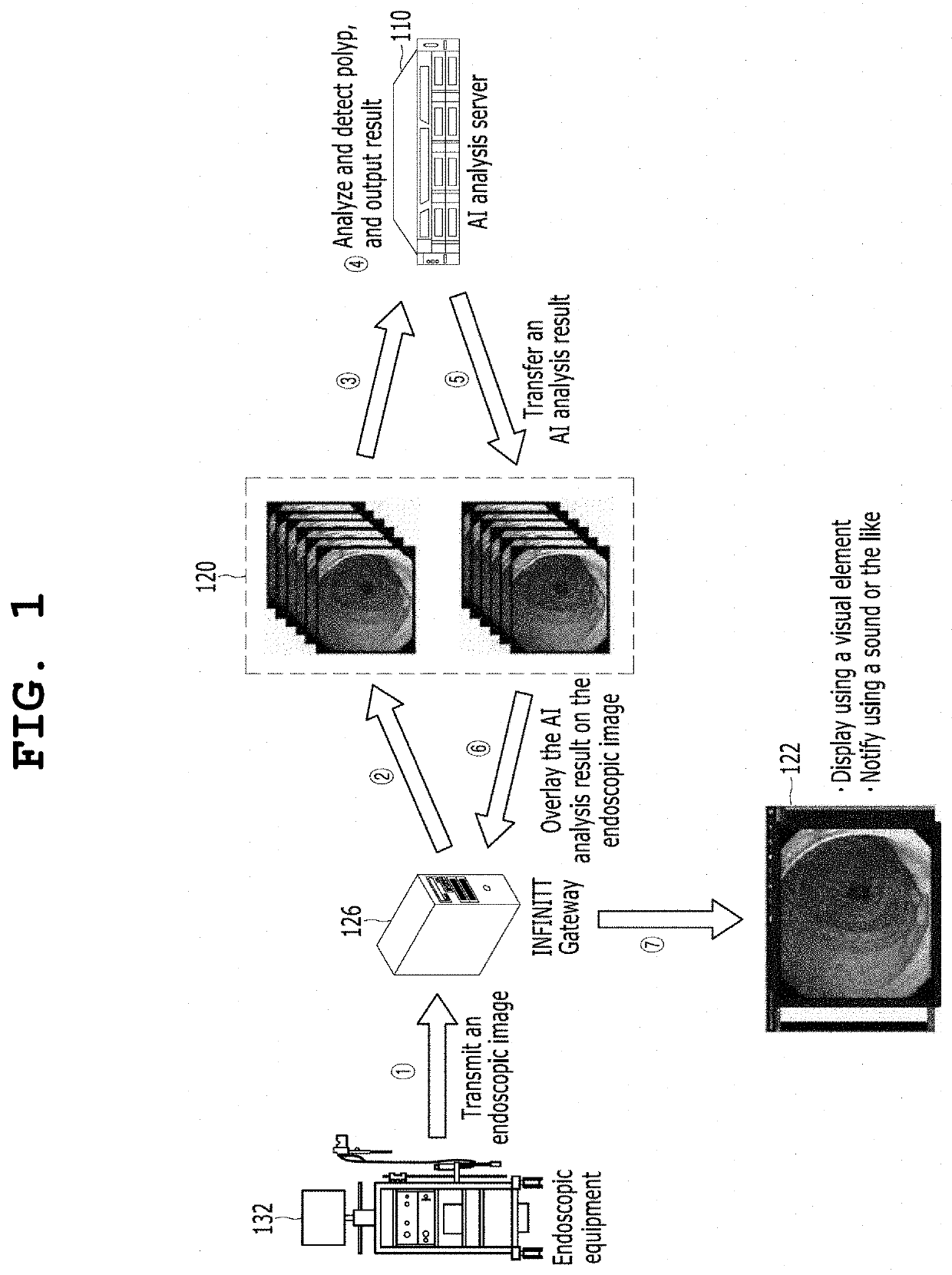
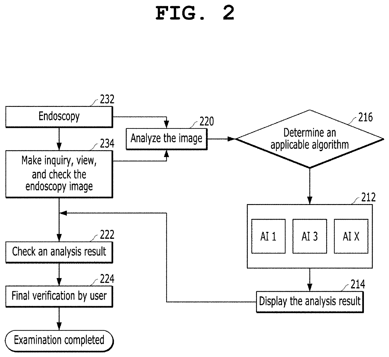
A system and method for assisting colonoscopic image diagnosis based on artificial intelligence include a processor, which is configured to analyze each video frame of a colonoscopic image using at least one medical image analysis algorithm and detects a finding suspected of being a lesion in the video frame. The processor calculates the coordinates of the location of the finding suspected of being a lesion. The processor generates display information, including whether the finding suspected of being a lesion is present and the coordinates of the location of the finding suspected of being a lesion.

Description

CROSS-REFERENCE TO RELATED APPLICATION[0001]This application claims priority from Korean Application No. 10-2021-0035619 filed on Mar. 19, 2021, which is incorporated herein by reference in its entirety.TECHNICAL FIELD[0002]The present invention relates to a medical image diagnosis assisting apparatus and method using an automated system. More particularly, the present invention relates to an apparatus, system, and method for assisting the diagnosis of a colonoscopic image during colonoscopy by using an artificial intelligence-based medical image analysis algorithm and also assisting the reduction of the risk of missing lesions.[0003]The present invention was derived from the research conducted as part of the SW Computing Industry Fundamental Technology Development Project sponsored by the Korean Ministry of Science and ICT and the Institute of Information & Communications Technology Planning & Evaluation [Project Serial No.: 1711116343; Project No.: 2018-0-00861-003; and Project Na...

Claims


Application Information

Patent Timeline
Patent Type & Authority Applications(United States)
IPC IPC(8): G06T7/00G06T7/70A61B1/31A61B1/00
CPCG06T7/0012G06T7/70A61B1/31A61B1/0005G06T2207/30028G06T2207/30096G06T2207/10016G06T2207/10068G06T2207/20084A61B1/000094A61B1/000096G06T2207/20081G06T7/0014G06T2207/20076G06T7/75G16H50/20G16H30/40G16H30/20G16H50/70A61B1/00011A61B1/00045A61B2576/00
Inventor BYUN, SOHYUNAHN, CHUNG IL
Owner INFINITT HEALTHCARE CO LTD
Learn More