Ultrasound image processing system

a processing system and ultrasound technology, applied in the field of ultrasound image processing system, can solve the problems of large amount of picture data, complicated data processing operations, and difficulty in obtaining information on the condition of internal structures or internal tissues of scanned subjects, and achieve the effect of large picture data to be processed

Inactive Publication Date: 2000-08-22
FUJI PHOTO OPTICAL CO LTD
View PDF5 Cites 47 Cited by
  • Summary
  • Abstract
  • Description
  • Claims
  • Application Information

AI Technical Summary

Problems solved by technology

Therefore, 3D ultrasound image processing normally involves a vast amount of picture data and involves extremely complicate data processing operations which take time even by a large-scale data processor.
From non-transparent 3D ultrasound images of this sort, it is difficult to obtain information on the conditions of internal structures or internal tissues of a scanned subject.
However, the greater the number of unit images, the larger becomes the amount of picture

Method used


Image

  • Ultrasound image processing system
  • Ultrasound image processing system
  • Ultrasound image processing system

Examples


Embodiment Construction

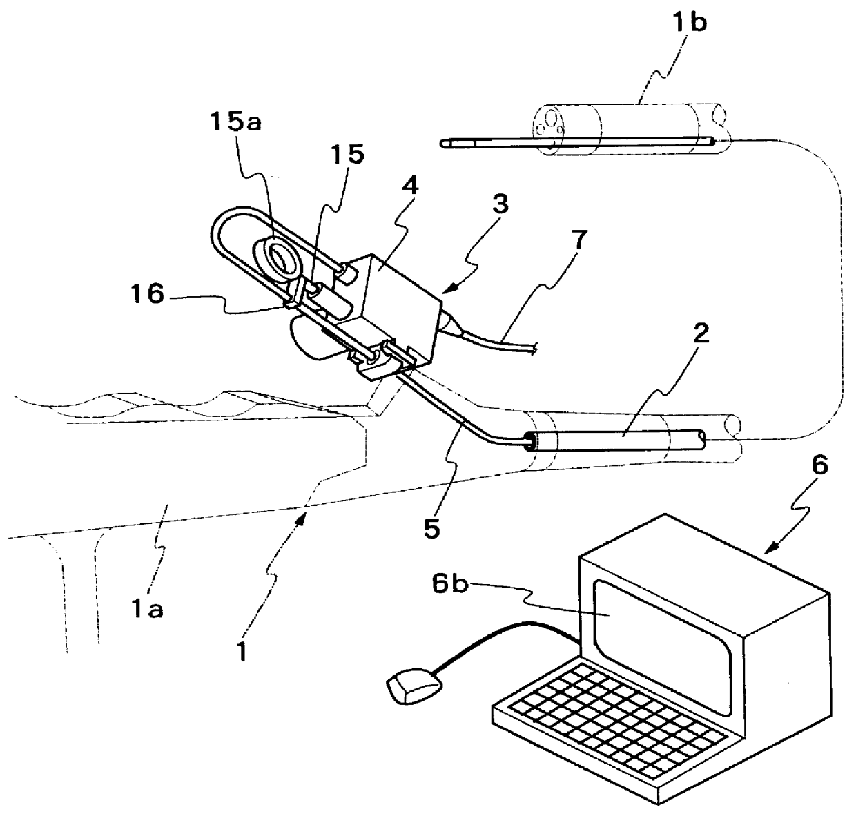
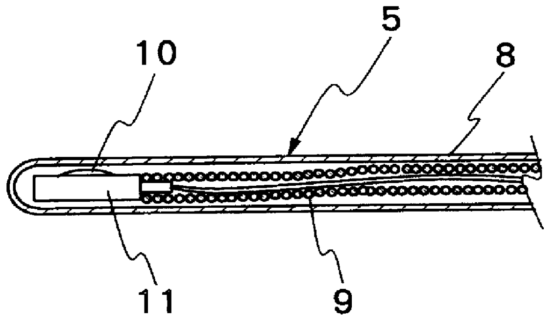
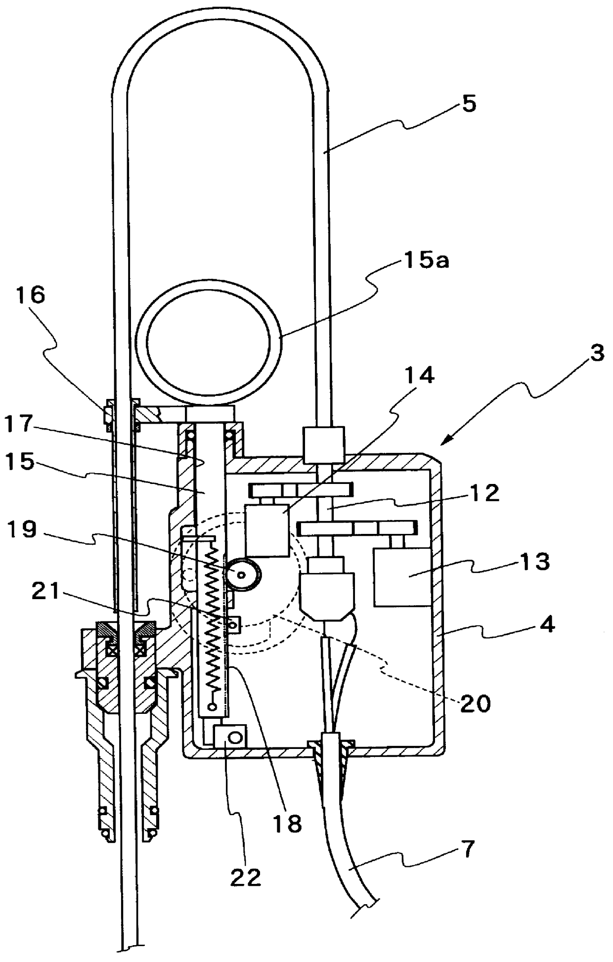
Hereafter, the invention is described more particularly by way of its preferred embodiment with reference to the accompanying drawings. FIGS. 1 to 3 shows, as one example of 2D ultrasound image capture means, an ultrasound examination system employing an endoscopically inserting ultrasound probe.

In FIG. 1, indicated at 1 is an endoscope which is largely constituted by a manipulating or gripping head 1a and an elongated insertion rod 1b extended out on the front side of the gripping head 1a for insertion into patient body. Extended through the endoscope 1, from a fore end portion of the gripping head 1a to the distal end of the insertion rod 1b, is a biopsy channel 2 for insertion of forceps or other bioptic instruments. For this purpose, the biopsy channel 2 is opened to the outside at the distal end of the insertion rod 1b. Indicated at 3 is an ultrasound probe which is mounted on the gripping head 1a of the endoscope 1, the ultrasound probe 3 having a manipulating head assembly 4 ...


PUM


Abstract

An ultrasound image processing system, having an ultrasound image capture device such as a B-mode ultrasound probe capable of making 2D scans over a predetermined range, and a drive for moving an ultrasound transducer in a direction different from the direction of 2D scans by the transducer to capture 2D ultrasound images sequentially in a predetermined pitch in the direction of movement of the ultrasound transducer. The ultrasound image processing system is provided with a marker for inserting a marker sign on field images selected from a large number of 2D ultrasound images sequentially captured by the image capture device, for use in three-dimensional image processing in a subsequent stage.

Description

1. Field of the ArtThis invention relates generally to an ultrasound image processing system for generating and displaying three-dimensional ultrasound images on the basis of a series of sequentially captured two-dimensional tomographic ultrasound images, and more particularly to an ultrasound image processing system having a marker means for marking and distinguishing two-dimensional ultrasound images, which are fundamentally necessary for three-dimensional image processing, from other 2D ultrasound images captured.2. Prior ArtIn ultrasound examination, ultrasound signals are transmitted into patient body through an ultrasound transducer, while thereby receiving return echo signals from body tissues at different depths in the directions of signal transmission. The return echo signals are processed into video signals to display ultrasound images on a monitor screen. A two-dimensional tomographic ultrasound image (hereinafter referred to as "2D ultrasound image" for brevity) is obtai...

Claims


Application Information

Patent Timeline
IPC IPC(8): A61B8/12A61B8/14G06T17/40G01B17/00A61B8/00G01N29/06G01N29/26
CPCA61B8/12A61B8/14A61B8/4209A61B8/463A61B8/483G01N29/0609G01N2291/02854
Inventor ISHIGURO, MASAAKI
Owner FUJI PHOTO OPTICAL CO LTD
Why Eureka
Try Eureka