Fluorescent imaging method of saccharide uptake activity of living tissue

a living tissue and fluorescence imaging technology, applied in the field of biochemistry, can solve the problems of insufficient spatial resolution, inability to allow, and no useful imaging methods for glucose uptake activity have been developed until now

Inactive Publication Date: 2001-03-27
NAT UNIV CORP TOKYO UNIV OF AGRI & TECH
View PDF0 Cites 29 Cited by
  • Summary
  • Abstract
  • Description
  • Claims
  • Application Information

AI Technical Summary

Problems solved by technology

However, no useful imaging methods for the glucose uptake activity has been developed until now.
However, this method does not allow, the bodies or tissues to be imaged as they are alive at real time, and requires complicated operations and is, furthermore, not sufficient in spatial resolution.
However, though these can image three-dimensionally, any of them have only a spatial resolution of about 0.1 mm, so that a resolution at a single cell level cannot be obtained.
Therefore, there still are many technical difficulties for obtaining images corresponding to the glucose uptake activity.
If the above reaction temperature is too low, the reaction rate will be low, requiring a long reaction time, while if it is too high, a fear of a side reaction or decomposition of reaction products will be posed.

Method used


Image

  • Fluorescent imaging method of saccharide uptake activity of living tissue
  • Fluorescent imaging method of saccharide uptake activity of living tissue
  • Fluorescent imaging method of saccharide uptake activity of living tissue

Examples


example 1

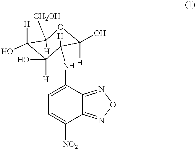
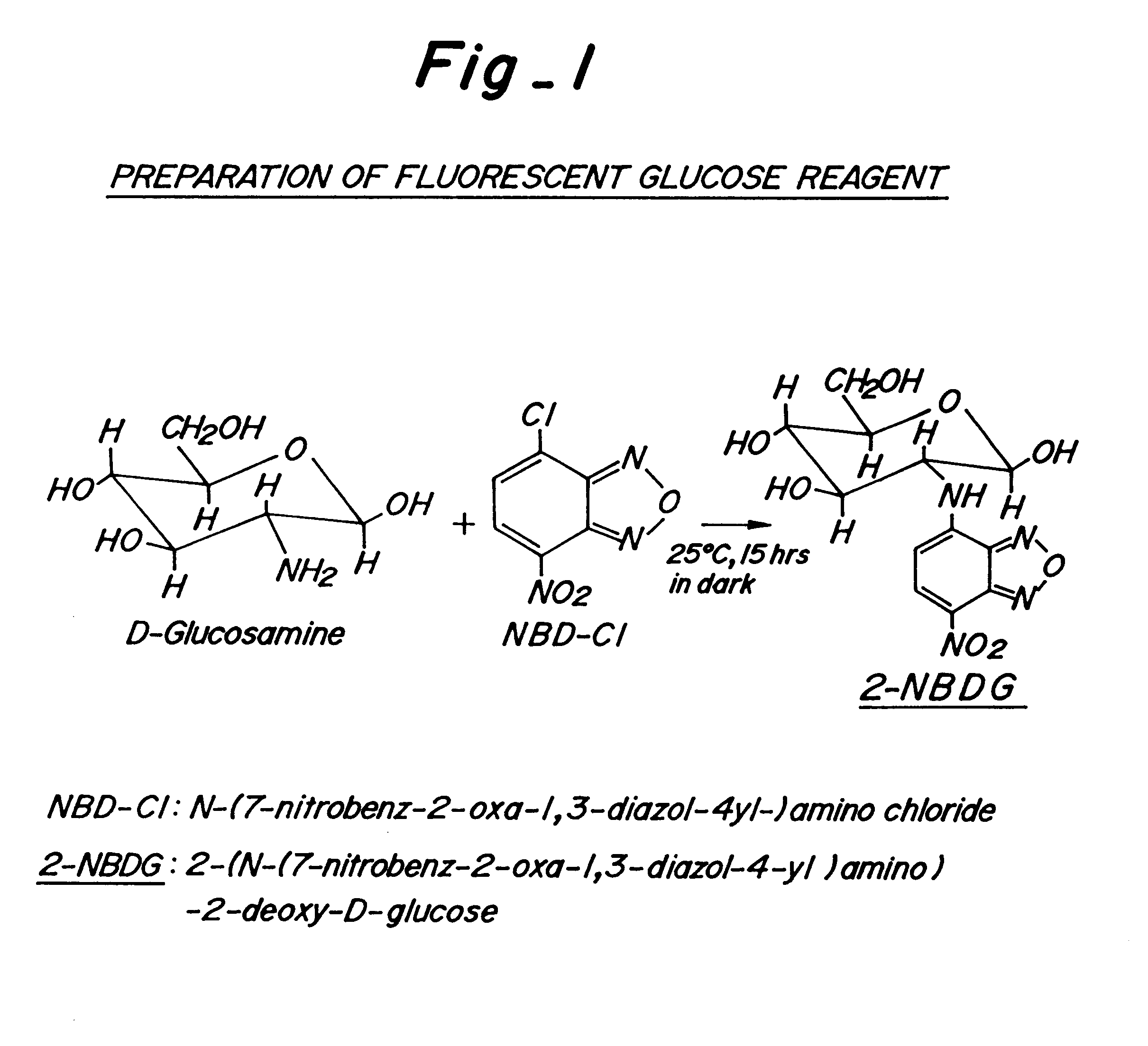
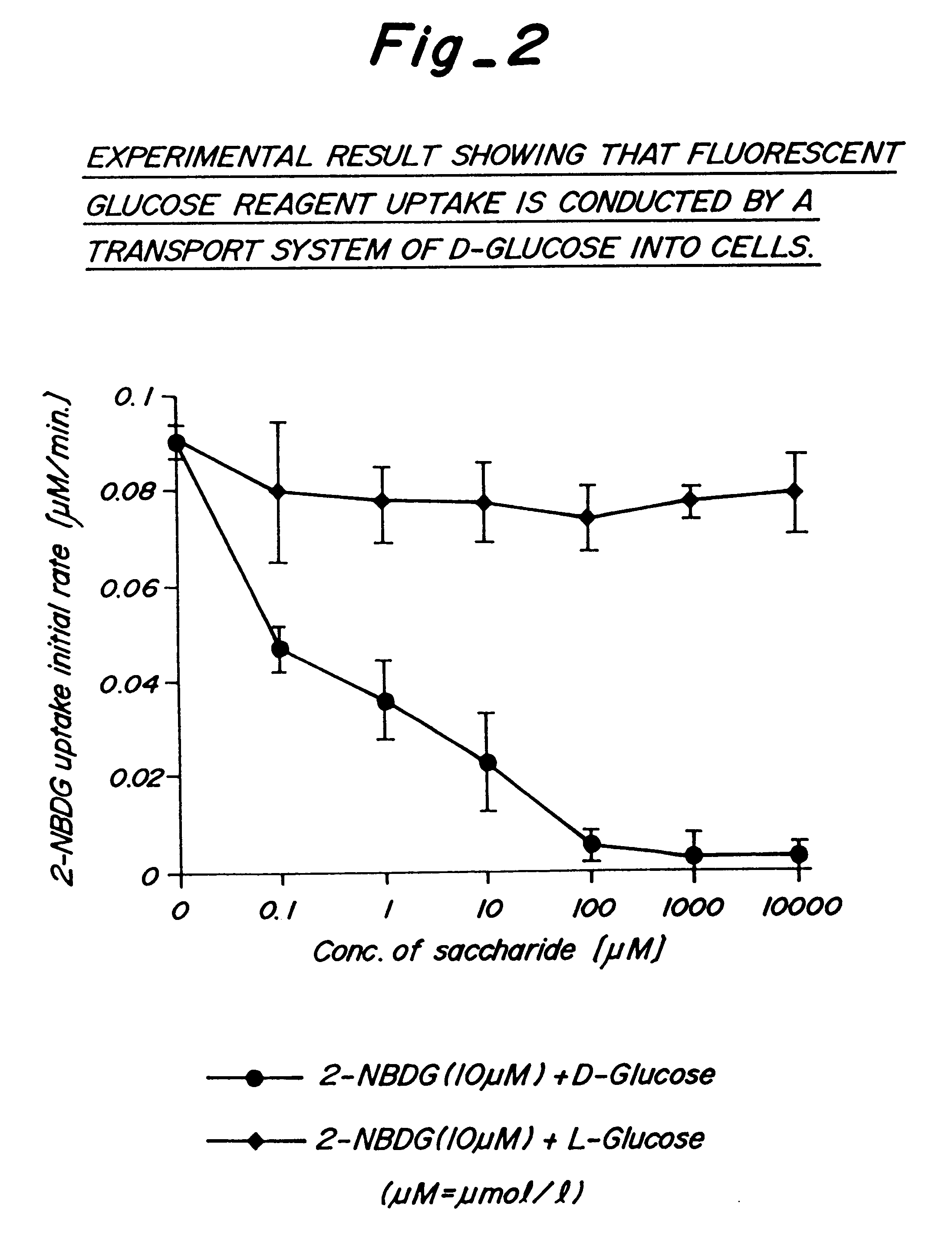
It was confirmed with cells of coliform bacillus, E. coli, and a yeast that the 2-NBDG (molecular weight: 342), which was a glucose derivative (molecular weight: 180) having at its C-2 position a side chain of molecular weight of at most 300, was incorporated equivalently to glucose. The graph shown in FIG. 2 is a result of the coliform bacillus. Namely, coliform bacilli (E. coli) were soaked in fluorescent saccharide reagents in which 10 .mu.M of the 2-NBDG coexisted with various concentrations of 0.about.10,000 .mu.M of D-glucose or L-glucose, and 2-NBDG uptake rates were investigated. As is apparent from the graph, the 2-NBDG uptake initial rate was substantially constant, irrespective of the concentration of L-glucose. However, when D-glucose coexisted, the uptake initial rate decreased rapidly with an increase in the D-glucose concentration, to substantially be near 0 when about a 10 fold concentration of the D-glucose coexisted. This suggested that the 2-NBDG uptake was compet...

example 2

The test for incorporating the 2-NBDG into living tissues taken out of animal or plant individuals can be performed in the same manner as with the microorganisms, such as E. coli or a yeast, in the above Example 1. In the case of the imaging of the glucose uptake activity of animal or plant individuals, setting up conditions were more difficult, and conducted as follows:

With respect to the brain of a silkworm, as shown in the illustrative chart of FIG. 3, 5th-instar silkworms were anesthetized by steeping in water for 30 minutes, and then 30 .mu.l of 10 mM 2-NBDG were injected into the head of the silkworms. The silkworms were divided into two groups: "odor stimulation group" in which the silkworm container received mulberry green leaves, and "control group" in which the silkworm container received no leaf. The brains of the silkworms of the two groups were extracted at every constant time interval, and observed with a confocal laser scanning fluorescence microscope. The results are...

example 3

The above example 2 showed imaging which was obtained as two-dimensional image information by direct continuous observation. However, if a confocal laser scanning fluorescence microscope is used, three-dimensional stereoscopic image information is also obtainable. The photomicrograph of FIG. 5 shows a 2-NBDG uptake activity after an hour of the brain of the silkworm shown in FIG. 4. In FIG. 5, as shown in (I) with dotted lines, in the brain tissues of the silkworm, the focal point of the confocal laser scanning fluorescence microscope to observe the brain tissues is slipped down successively by 5 .mu.m, whereby optical slices A, B, C, D, E and F are obtained. The images taken from the above within the frame (II) were shown in photomicrographs A, B, C, D, E and F, respectively, in FIG. 5. By the thus obtained sectional imaging, stereoscopic image information is continuously obtained with a spatial resolution of about 1 .mu.m as the living tissues, etc. maintain physiological activity...


PUM

PropertyMeasurementUnit
temperatureaaaaaaaaaa
molecular weightaaaaaaaaaa

Abstract

A fluorescent imaging method of the changes in viability of living bodies wherein the living bodies are provided with a chemical or physical exterior stimulation and information on the changes in viability of cells caused by the stimulation is obtained by means of a fluorescent imaging method of the saccharide uptake activity by contacting or administering to the living bodies an aqueous solution of a fluorescent saccharide compound which includes at its specific position a saccharide molecule chemically combined with a fluorescent pigment molecule.

Description

1. Field of the InventionThe present invention, in the field of biochemistry, particularly relates to a method for imaging the saccharide uptake activity of living cells which are in a state of maintaining biological activity and for obtaining information relating to changes in viability of living cells with external stimulations.2. Description of the Prior ArtIt is considered that the most universal indicator for representing the viability of living cells is an energy source uptake activity (uptake rate or uptake quantity). The typical example what of many of those cells can commonly take up as an energy source is glucose. However, no useful imaging methods for the glucose uptake activity has been developed until now.Heretofore, as a method for imaging the glucose uptake activity in living bodies or tissues thereof, a 2-deoxyglucose (2-DG) method has been conducted. Namely, it is a method in that after a radioisotope labeled 2-deoxyglucose is incorporated instead of glucose, slices...

Claims


Application Information

Patent Timeline
Patent Type & Authority Patents(United States)
IPC IPC(8): C07H5/06C07H5/00C07H15/00C07H15/26A61B5/00A61B10/00A61K49/00G01N21/64C07D271/08C07H17/00C09K11/06G01N33/48
CPCC07H5/06C07H15/26A61B5/14532A61B5/1455
Inventor MATSUOKA, HIDEAKI
Owner NAT UNIV CORP TOKYO UNIV OF AGRI & TECH
Why Eureka
Try Eureka