Method for transdermal sampling of analytes

Inactive Publication Date: 2002-05-07
HEALTH RES INC
View PDF8 Cites 18 Cited by
  • Summary
  • Abstract
  • Description
  • Claims
  • Application Information

AI Technical Summary

Problems solved by technology

However, pain and inconvenience associated with frequent drawing of blood may cause patients to miss monitoring routines.
However, the non-permeable nature of SC and the side effects associated with iontophoresis such as irreversible skin damage, limit the use of iontophoresis for transdermal sampling.
However,.the impermeability of the stratum corneum layer l

Method used


Image

  • Method for transdermal sampling of analytes
  • Method for transdermal sampling of analytes
  • Method for transdermal sampling of analytes

Examples


example 2

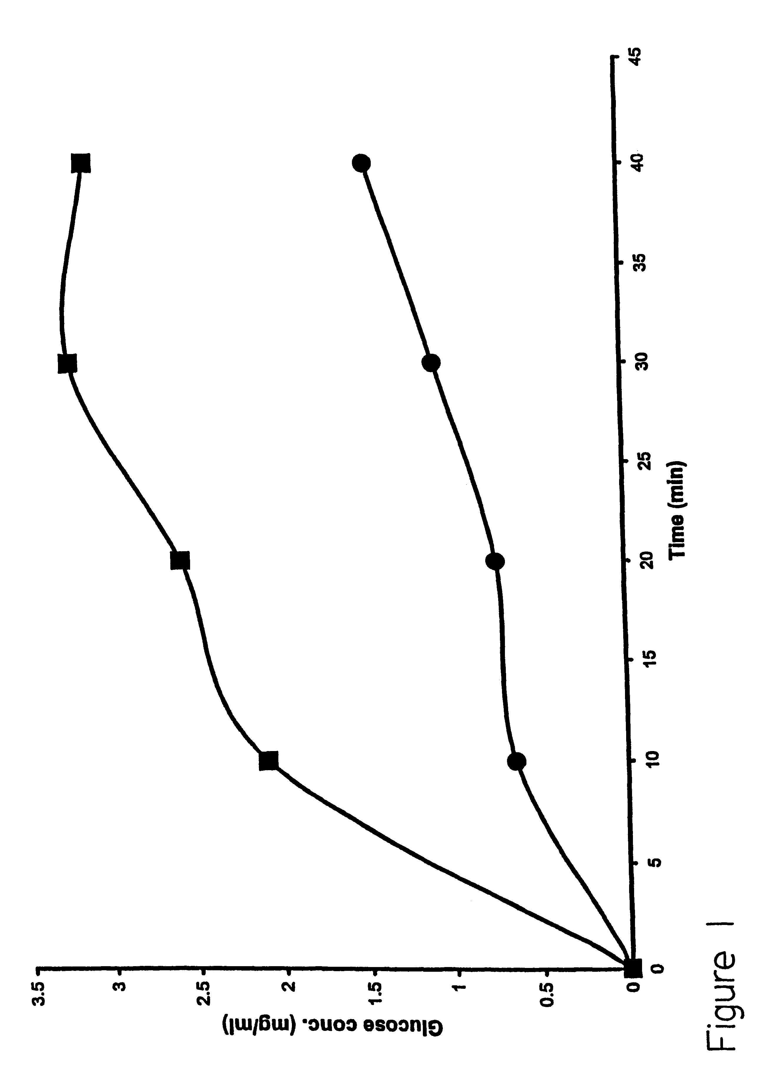
This embodiment illustrates that the analyte glucose can cross the stratum corneum using the method of the present invention. The stratum corneum layer was obtained by heat treatment of porcine skin-as-follows. Fresh pieces of porcine skin were wrapped in aluminum foil and placed in a 60.degree. C. water. In about 5 minutes, the stratum corneum can be gently pulled away by hand from the rest of the skin tissue. The isolated SC layer was used in a Vertical Diffusion Chamber (such as commercially available from Crown Glass Co., Sommerville, N.J.). This simple device is well accepted as a model system for skin. The upper chamber was considered as the outer surface of the skin and the lower chamber as the skin directly below the stratum corneum. Platinum wires served as electrodes, one was placed in the upper chamber and the other in the lower chamber. Electric pulses were applied across the SC using a pulse generator

The lipid formulation prepared as described in Example 1 was placed in...

example 3

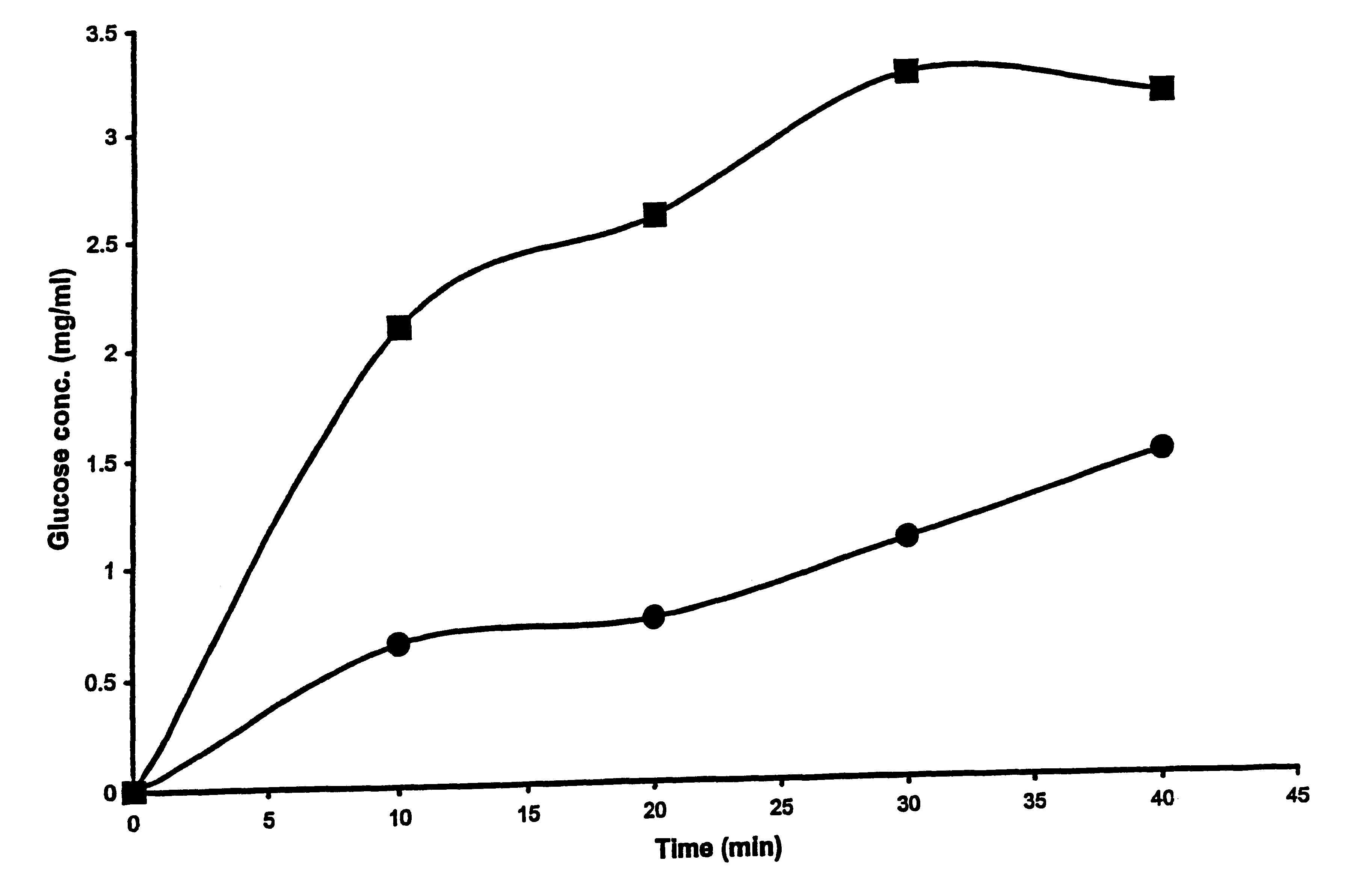
This embodiment illustrates that the presence of liposomes enhances the permeability of whole skin during application of electric pulses. To illustrate this embodiment, freshly excised, full thickness mouse skin was used as a model to investigate the permeability changes following application of varying numbers of electric pulse with and without liposomes. Electrical resistance of the skin was measured as an indicator of skin permeability. The drop in the skin resistance with electric pulse application and the subsequent recovery of the resistance with time after end of pulse application was measured. The time during which the skin resistance remained lower than its base value (before any electric pulse was applied) was taken as the time the skin remained permeable due to electric pulse application.

Electric pulse were applied as described earlier, using a vertical diffusion chamber. The excised full thickness mouse skin was placed between the two aqueous compartments as a barrier. B...

example 4

This embodiment illustrates that the method of the present invention enhances the extraction of glucose in whole animals. Systemic glucose levels in laboratory mice (Strain CD-1, supplier Charles River) were measured by applying electric pulse to bare skin surface on the backs of the animals using a pair of commercially available electrodes. The electrode assembly consists of an Ag / AgCl electrode inside a small reservoir cup (1 cm diameter, 5 mm deep). The cup was filled with cotton wool soaked in buffer alone or buffer with added liposomes. Electrical contact between the electrodes and the skin was established via the buffer-soaked cotton wool. The mice were anesthetized and hair was clipped from backs of the animals to expose bare skin. The mice were kept under anesthesia during the whole procedure. The electrode assemblies were placed on backs of the animals and taped in place. One of the electrodes was maintained at ground potential and negative pulses (.sup..about. 200V, 1 ms p...


PUM


Abstract

The present invention discloses a method for transdermal extraction and measurement of analytes in an individual's body comprising the steps of permeabilizing the skin by electroporation in the presence of liposomes comprising negatively charged lipids, and extracting the extracellular fluid through the permeabilized skin by using suction. Analytes may be quantitated in the extracted extracellular fluid.

Description

The present invention relates generally to the field of transdermal extraction and sampling of analytes. More particularly, the present invention provides a method for extraction and sampling of analytes by electroporation in the presence of liposomes.DISCUSSION OF RELATED ARTThe ability to accurately measure analytes such as glucose in the blood is important for management of disease. For example, proper monitoring of blood glucose levels prior to or following insulin injections could result in easier management of diabetes.Conventional monitoring of blood analytes requires drawing of a blood sample. However, pain and inconvenience associated with frequent drawing of blood may cause patients to miss monitoring routines. To overcome problems associated with invasive techniques, several approaches have been proposed that involve extraction of analytes through skin.Mammalian skin has two layers, the epidermis and the dermis. The epidermis is a stratified squamous keratinizing epitheli...

Claims


Application Information

Patent Timeline
IPC IPC(8): A61B5/00A61B5/145G01N33/48A61B5/15A61B5/157C12Q1/54G01N33/66
CPCA61B5/14514A61B5/14546A61B5/14532A61M2037/0007
Inventor SEN, ARINDAMHUI, SEK WENZHAO, YA LI
Owner HEALTH RES INC
Why Eureka
Try Eureka