Ultrasound imaging with acquisition of imaging data in perpendicular scan planes

Inactive Publication Date: 2003-03-25
SIEMENS MEDICAL SOLUTIONS USA INC
View PDF10 Cites 91 Cited by
  • Summary
  • Abstract
  • Description
  • Claims
  • Application Information

AI Technical Summary

Problems solved by technology

This reduces the desirability of intracardiac imaging transducers, which are carried at the end of a catheter that is threaded into the heart via a major vein in the arm, neck or groin.
Unfortunately, the complexity and cost of such a real-time 3-D (sometimes referred to as 4-D) system are formidable.
Unfortunately, the element count for such a 2-D array is usually much larger (typically, well over 4000 elements) than for a 64-element 1-D array in order to achieve the same imaging performance.
Even with these techniques, a sparsely sampled aperture still requires a significant number of elements, typically at least ten times greater than a conventional 64-element array.
Because of the large number of elements that must be addressed in a sparse 2-D array, multiplexing (synthetic aperture) is sometimes used, but this reduces the frame rate.
This design has several weaknesses, among which are the increased difficulty of providing signal leads to the individual elements, even those that are to be connected to each other and/or decreased dicing pitch, which is the distance between the lines separating the elements.
Because the electrical connections will have to "hop over" every other element, the interconnection scheme is greatly complicated, espec

Method used


Image

  • Ultrasound imaging with acquisition of imaging data in perpendicular scan planes
  • Ultrasound imaging with acquisition of imaging data in perpendicular scan planes
  • Ultrasound imaging with acquisition of imaging data in perpendicular scan planes

Examples


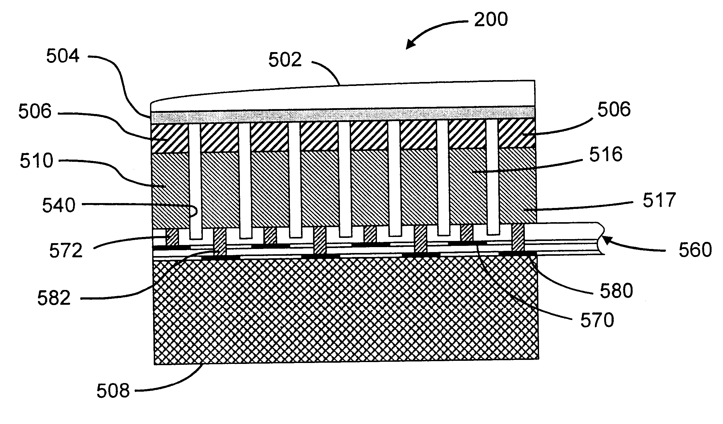
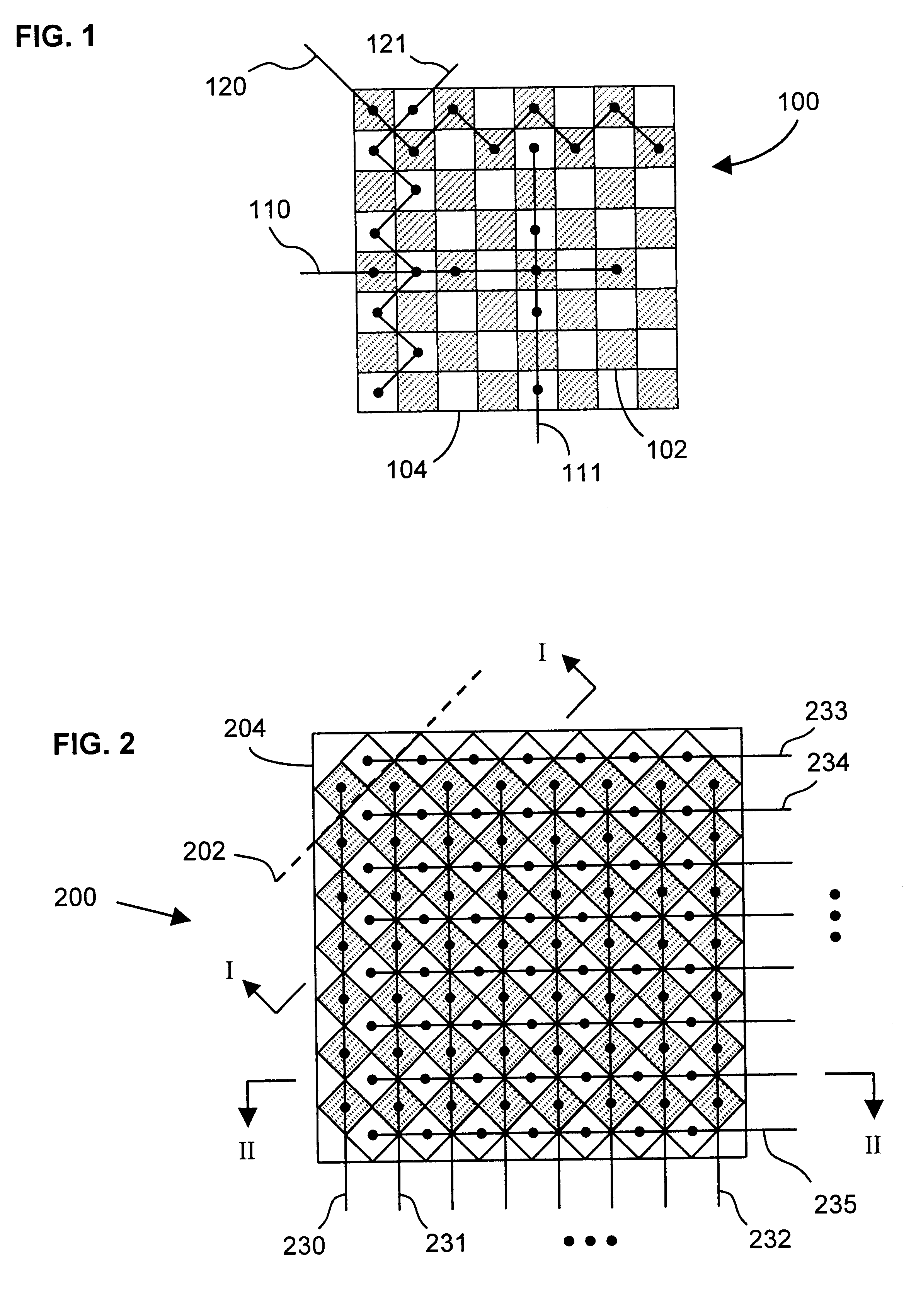
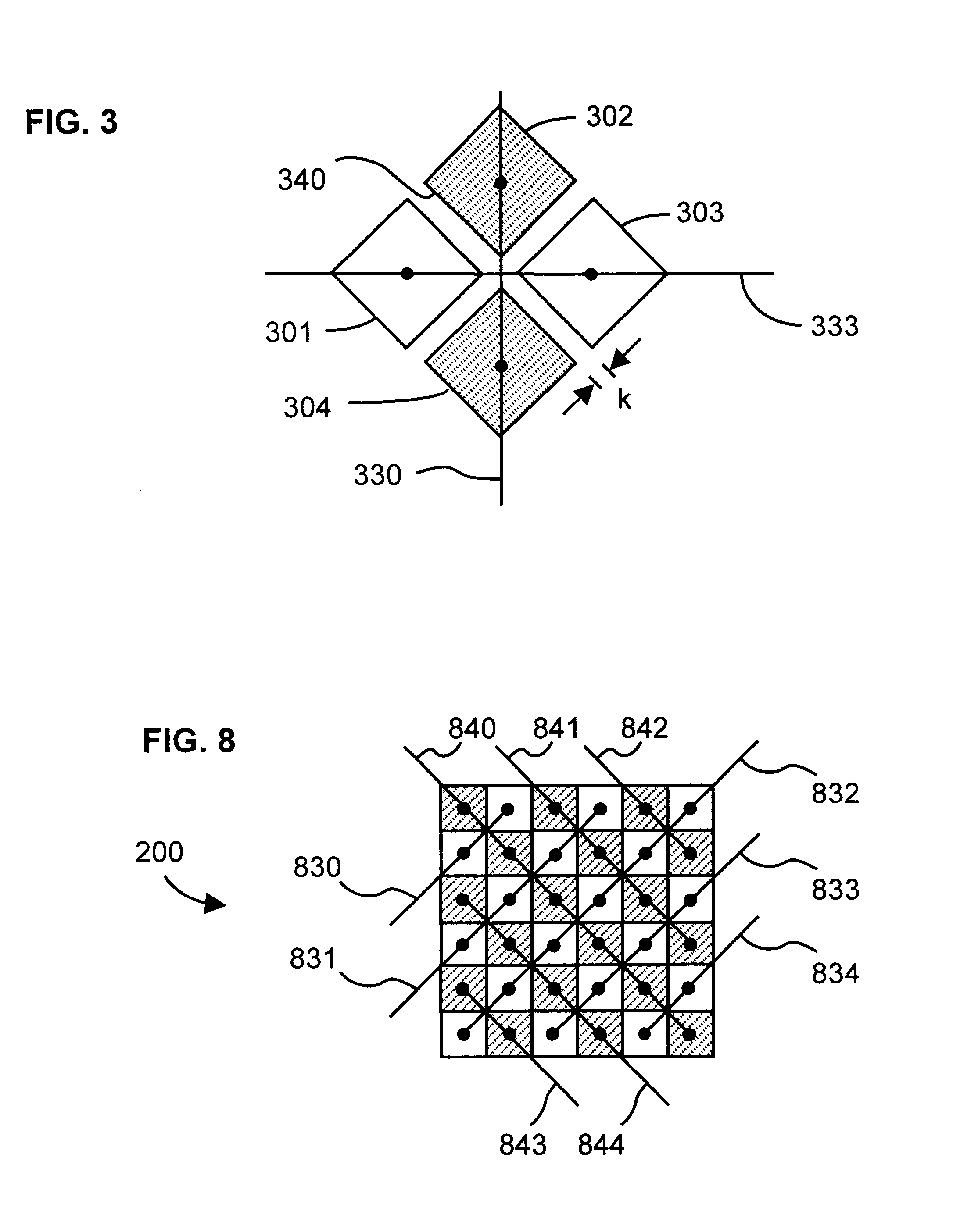
Embodiment Construction

The lens 520 in the preferred embodiment of the invention is designed to improve the ability of the array to independently and simultaneously adjust the focus of the two apertures, that is, of the two orthogonal scan planes made possible by the invention. In order to better understand the lens used in the invention, it is helpful to recall the way in which conventional arrays focus. These methods are therefore reviewed here.

In conventional linear arrays, there are two primary methodologies for mechanically focusing the array in elevation where electronic delay control does not exist. According to the first method, the geometric focus is created by curving the ceramic. Typically, this is done by using a convex-concave ceramic as shown in FIG. 13A. The geometric focus is simply given by the following equation: ##EQU2##

where the radius of curvature of the ceramic is equal to the geometric focus in elevation. This curvature only occurs in one dimension for a conventional linear array.

Th...


PUM


Abstract

An ultrasonic imaging array has two independent, interleaved linear subarrays occupying a common array face. The subarrays are independently steerable and focusable in different imaging planes. The subelements making up the elements in of the subarrays is preferably quadrilateral. Adjacent subelements in each element are electrically connected via an interconnect portion such that the interconnect portion connecting each pair of adjacent subelements in each element is substantially linear and is aligned with the diagonals of the adjacent subelements. A preferred acoustic lens is curved in both azimuth and elevation directions, whereby the subarrays are independently focusable. By transmitting in one plane and simultaneously receiving in a different, orthogonal plane, a system including the array can image a 3-D region of interest (ROI). In a volumetric embodiment of the invention two orthogonal B-mode images of a structure within the ROI are displayed simultaneously. The user may then trace the outlines of the images and the system therefrom calculates the volume of the structure.

Description

1. Field of the InventionThis invention relates in general to an ultrasound transducer that has a two-dimensional (2-D) array of transducer elements, and in particular to a 2-D transducer array for acquiring imaging data in different scan planes.2. Description of the Related ArtThe value of ultrasonic imaging as a diagnostic tool is undisputed, and as the power of ultrasound imaging systems increases, so too do the number of applications of the technology. Examples of such applications include the determination of the size and location of tumors and arterial occlusions. Another application that shows growing promise is in the area of cardiac imaging. As in many areas of diagnostics, however, physicians (and of course, patients themselves) prefer minimally, or, ideally, completely non-invasive techniques. This reduces the desirability of intracardiac imaging transducers, which are carried at the end of a catheter that is threaded into the heart via a major vein in the arm, neck or gr...

Claims


Application Information

Patent Timeline
IPC IPC(8): A61B8/14B06B1/06G01S15/00G01S15/89G10K11/30G10K11/00
CPCA61B8/14A61B8/145A61B8/4483G10K11/30G01S15/8925G01S15/8927G01S15/8993B06B1/0629
Inventor FRIEMEL, BARRY HUGHEMERY, CHARLES DALE
Owner SIEMENS MEDICAL SOLUTIONS USA INC
Why Eureka
Try Eureka