Method of diagnosing autoimmune disease

a technology of autoimmune disease and diagnosis method, which is applied in the field of diagnosis of autoimmune disease, can solve the problems of large discomfort and pain in patients, inability to find a patient, and limited criteria

Inactive Publication Date: 2003-07-22
FRED HUTCHINSON CANCER RES CENT
View PDF16 Cites 70 Cited by
  • Summary
  • Abstract
  • Description
  • Claims
  • Application Information

AI Technical Summary

Problems solved by technology

Thus, despite the presence of such antibodies, a clear diagnostic assay for individuals with SLE is not available due to the heterogeneous nature of SLE and other autoimmune diseases, requiring the diagnosis to be based on an array of different criteria.
Furthermore, these criteria are lim

Method used


Image

  • Method of diagnosing autoimmune disease
  • Method of diagnosing autoimmune disease
  • Method of diagnosing autoimmune disease

Examples


example 1

Materials & Methods

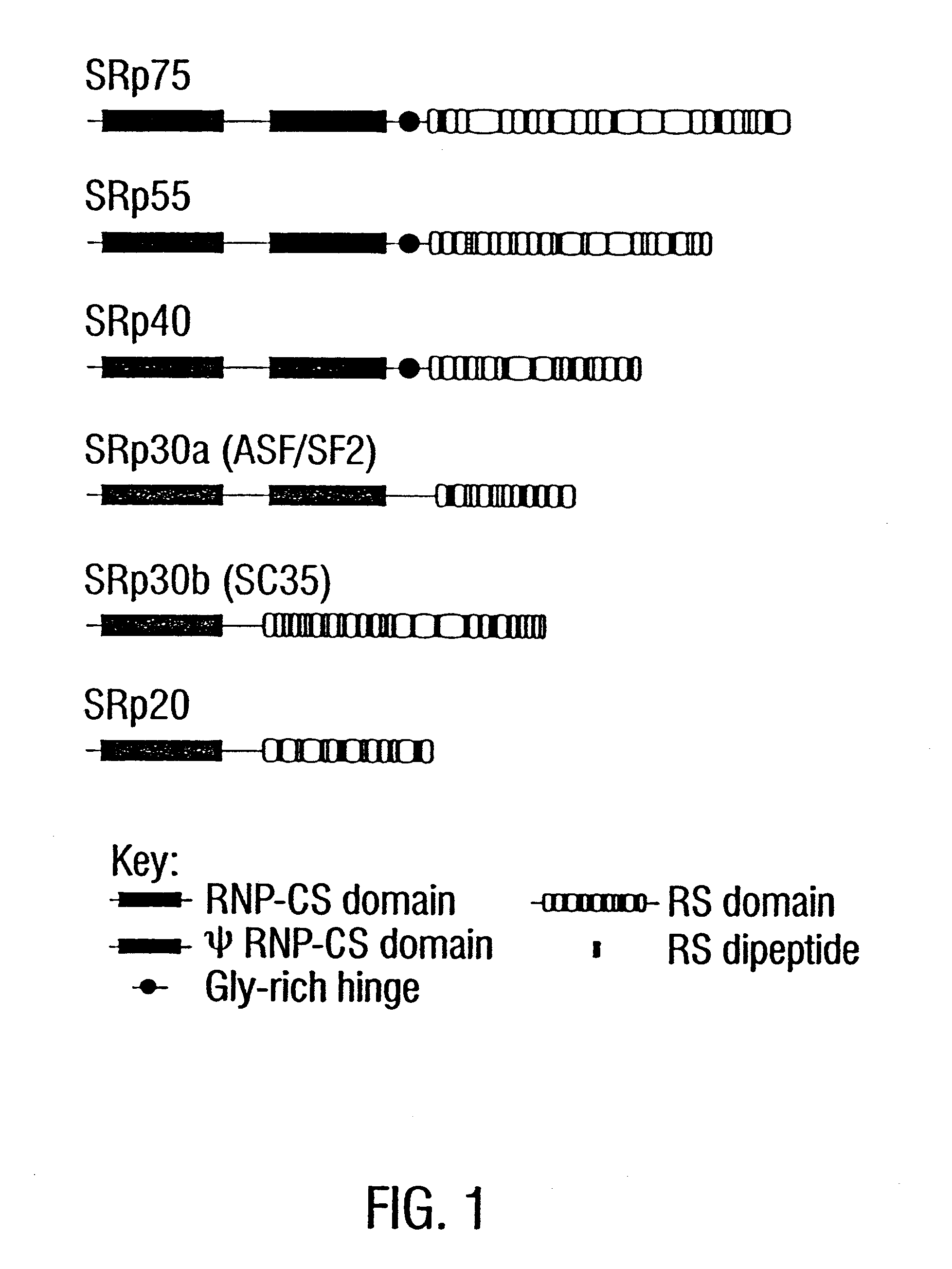
A. Purification and Microsequencing of SR Proteins

Protein purifications were carried out as described in Zahler et al., 1992, and are incorporated herein by reference. The purification was started with 1.times.10.sup.10 HeLa cells or 100 grams of calf thymus. Cells and tissues were ground to a fine powder in Liquid N.sub.2 using a mortar and pestle. The powder was then transferred to a beaker on ice, and 350 ml of isolation buffer was added (isolation buffer is 65 mM KCl, 15 MM NaCl, 10 mM HEPES at pH 7.6, 10 mM Na.sub.2 EDTA, 5 mM potassium fluoride (KF), 5 mM .beta.-glycerophosphate, 0.2 mM PMSF, and 2 .mu.g / ml of aprotinin). Tissue culture cells were sonicated 10 times for 20 sec with a probe sonicator set at 50 W; thymus was sonicated continuously for 20 min. Homogenates were centrifuged for 20 min at 8000.times.g, supernatants were transferred to a beaker, and ammonium sulfate was added to 65% of saturation. After 2 h of stirring at 4.degree. C., the extract ...

example 2

Results & Discussion

Results

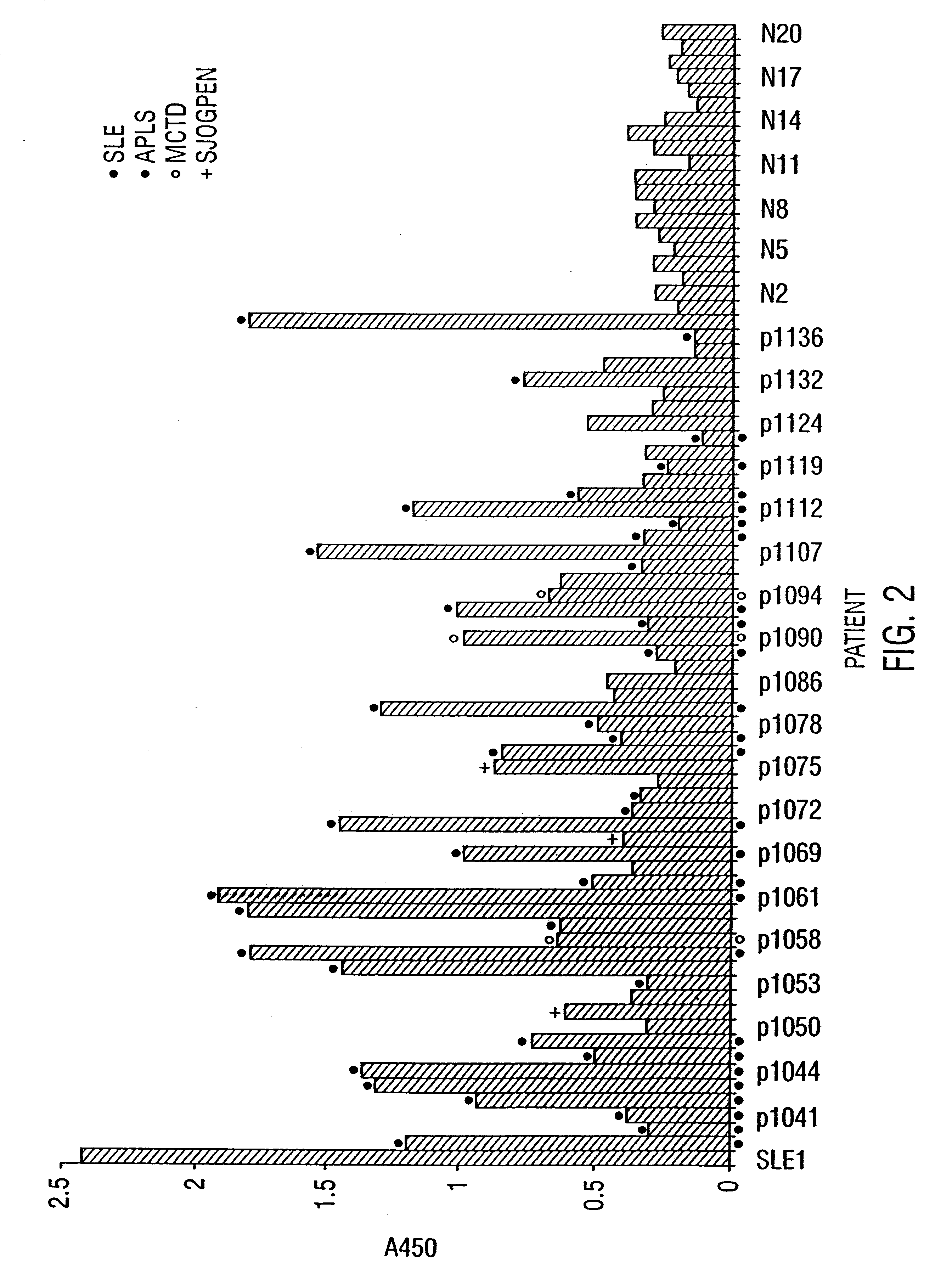
To determine whether SR proteins are autoantigens in any of a variety of diseases, an ELISA assay was developed. Human SR proteins were purified to homogeneity from HeLa cells grown in suspension and used to coat immunoplates at concentrations ranging from 0.5 to 2 mg / ml. At these coating concentrations, a monoclonal antibody (mAb) specific for the SR protein family, mAb1H4 (Neugebauer and Roth, 1997), gives a robust signal in the ELISA assay. In contrast, mAb Y12 against the Sm epitope gives no signal above background, indicating that the SR protein preparation is not detectably contaminated with snRNPs.

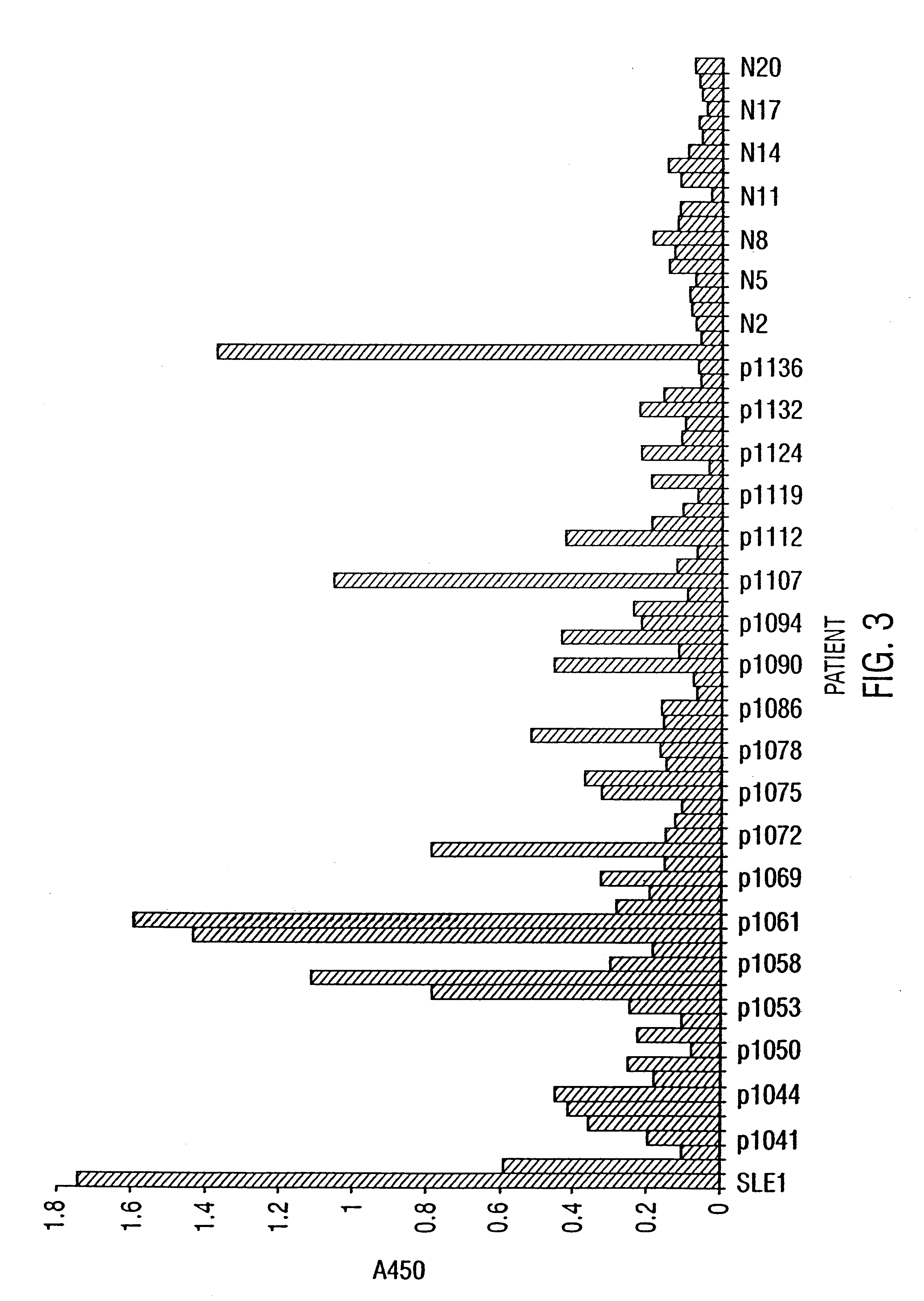
The inventor performed an ELISA assay for SR protein reactivity on sera obtained from patients with SLE, APLS, MCTD, PSS and Sjorgens syndrome. FIG. 2 and FIG. 3 show the presence of autoantisera against SR proteins at a 1:50 dilution and 1:200 dilution respectively. These data provide clear evidence of the presence of anti-SR protein antibodies in patient...


PUM


Abstract

The present invention relates to diagnostic applications. For autoimmune diseases more particularly, it is demonstrated herein that individuals with SLE, APLA, MCDS and PSS have antibodies that are specific for SR proteins. Thus, in particular aspects the present invention provides methods and compositions for diagnosing autoimmune disease using SR proteins and antibodies to detect the presence of SR protein-specific antibodies in an individual suspected of having autoimmune disease, wherein the presence of such antibodies is indicative of said individual suffering from autoimmune disease.

Description

1. Field of the InventionThe present invention relates generally to the field of diagnosing autoimmune disease. More particularly, it concerns the use of specific antigens to detect the presence of SR protein-specific antibodies in an individual suspected of having autoimmune disease, wherein the presence of such antibodies is indicative of said individual suffering from autoimmune disease.2. Description of Related ArtAutoimmune diseases are known to afflict a significant portion of the population. Some of the more common autoimmune diseases include, among others, scleroderma, systemic lupus erythrematosus and mixed connective tissue disease. These diseases are characterized by the presence of a multitude of autoreactive antibodies that arise spontaneously. To date, high levels of circulating autoantibodies to DNA are the best evidence of these maladies.In the blood of systemic lupus erythematosus (SLE) patients, there typically are found antibodies directed against one or more comp...

Claims


Application Information

Patent Timeline
IPC IPC(8): G01N33/564
CPCG01N33/564G01N2800/24Y10S977/959
Inventor ROTH, MARK
Owner FRED HUTCHINSON CANCER RES CENT
Why Eureka
Try Eureka