Sensitive and rapid methods of using chimeric receptors to identify autoimmune disease and assess disease severity

a technology of chimeric receptors and autoimmune diseases, applied in the field of diagnostics of autoimmune diseases, can solve the problems of cumbersome and laborious commercial methods, poor diagnostic accuracy, and inability to accurately predict the severity of the disease, and achieve the effect of high predictive valu

Inactive Publication Date: 2013-10-22
DIAGNOSTIC HYBRIDS
View PDF4 Cites 7 Cited by
  • Summary
  • Abstract
  • Description
  • Claims
  • Application Information

AI Technical Summary

Problems solved by technology

Graves' disease (also referred to as “diffuse toxic goiter”), is the leading cause of hyperthyroidism due to the action of autoantibodies that recognize and bind to receptors present on the thyroid gland, resulting in gland growth and over-production of thyroid hormone.
Current diagnostic techniques for Graves' disease leave much to be desired.
In general, the commercially available methods are cumbersome and laborious.
Most importantly, however, the vast majority of the presently used methods lack sufficient sensitivity such that a quick, accurate and cost-effective test can be performed.

Method used


Image

  • Sensitive and rapid methods of using chimeric receptors to identify autoimmune disease and assess disease severity
  • Sensitive and rapid methods of using chimeric receptors to identify autoimmune disease and assess disease severity
  • Sensitive and rapid methods of using chimeric receptors to identify autoimmune disease and assess disease severity

Examples


example 1

Preparation of Cho-Rluc Cells for Testing

[0209]In these experiments, CHO-Rluc cells were prepared from W-25 CHO-R cells for use in the testing methods to detect TSI in Graves' disease patients. Pools of puromycin-resistant cells were obtained and tested for light output in response to bovine TSH. Clones with the highest light output were selected for use in the experiments described below.

[0210]CHO-Rluc cells were grown in cell culture flasks (e.g., T-225 flasks) in growth medium containing Ham's F-12 medium, 10% FBS (heated at 56° C. for 30 minutes to inactivate complement), 2 mM glutamine, and 1× non-essential amino acids. The flasks were incubated at 35-37° C., in a humidified atmosphere, containing 5% carbon dioxide.

[0211]After the cell cultures reached confluence, the medium from each flask was aspirated, and the cell monolayers were washed with HBSS without Ca and Mg++. Then, 7 ml of a 0.25% trypsin / 1 mM EDTA solution were added to each flask, and allowed to react with the mon...

example 2

CHO-Rluc Assay Plate Preparation and Testing

[0213]In these experiments, CHO-Rluc cells prepared as described in Example 1 were used in assays for diagnosis of Graves' disease. To prepare 24 monolayers for testing, 24 wells in a 96-well microtiter plate were first treated by adding 50-100 μl 0.1% gelatin solution (Sigma) to enhance attachment of the cells to the bottom of the 24 wells chosen for the test. Following incubation for approximately 1 minute at room temperature, the gelatin solution was removed from each of the wells by aspiration. It was noted that the gelatin can remain on the wells for longer than one minute. The gelatin serves to coat the wells with collagen, so that the cells attach more quickly to the wells and reach confluence more rapidly. However, cells can be planted and grown to confluence without gelatin and still perform well.

[0214]A freezer vial of CHO-Rluc cells produced as described in Example 1 was rapidly thawed in a 37° C. water bath to provide approxima...

example 3

Preparation of IgG Samples

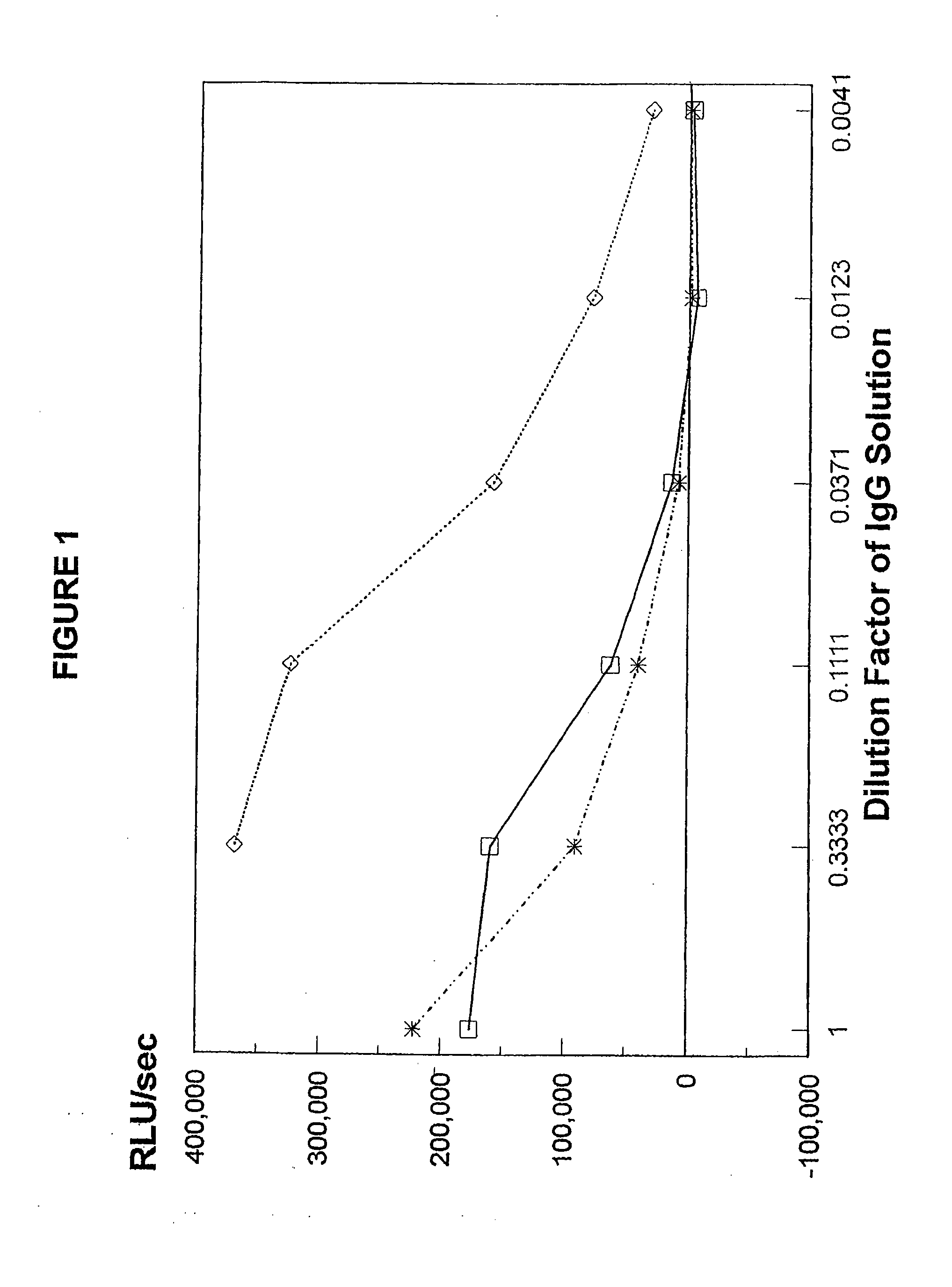
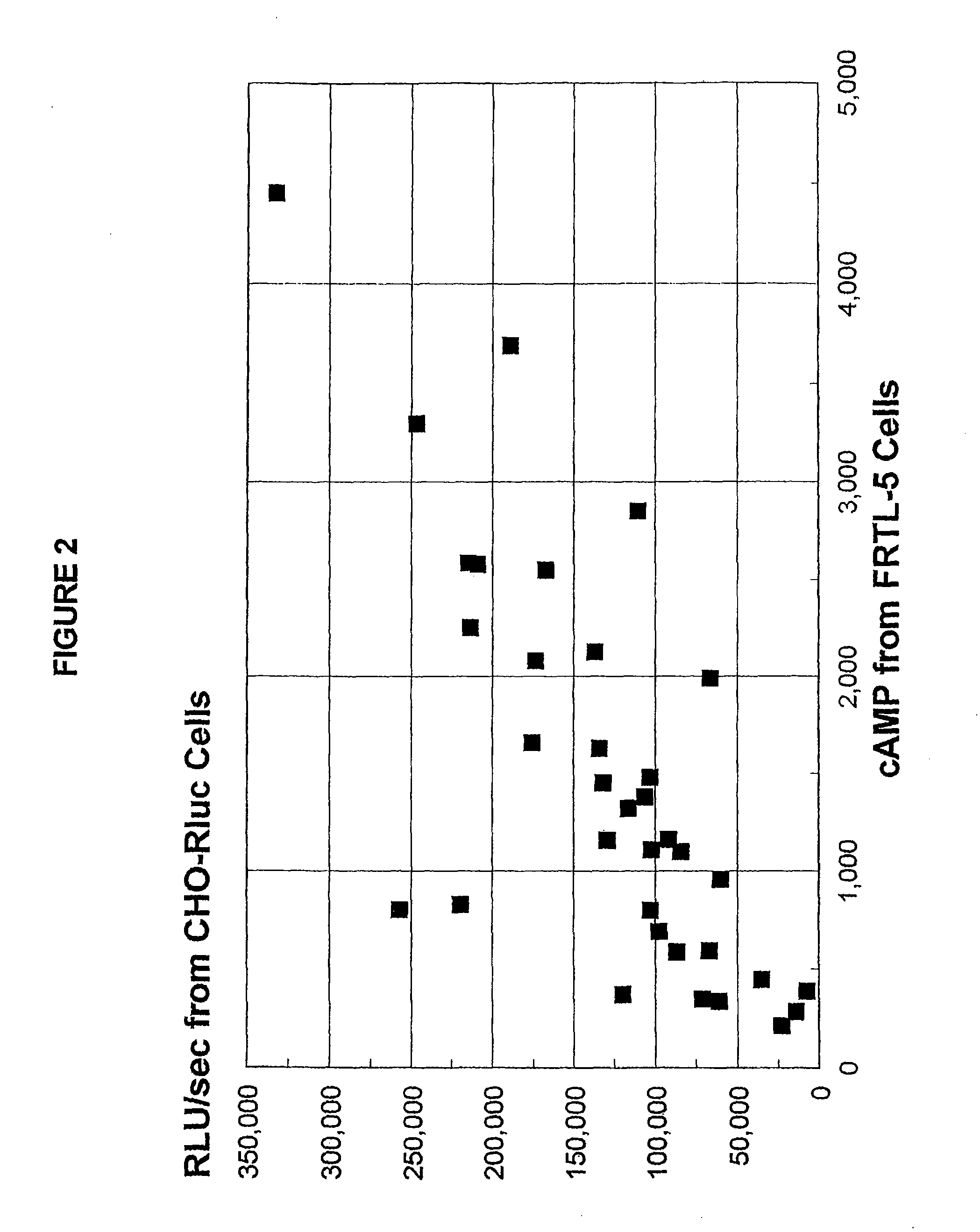
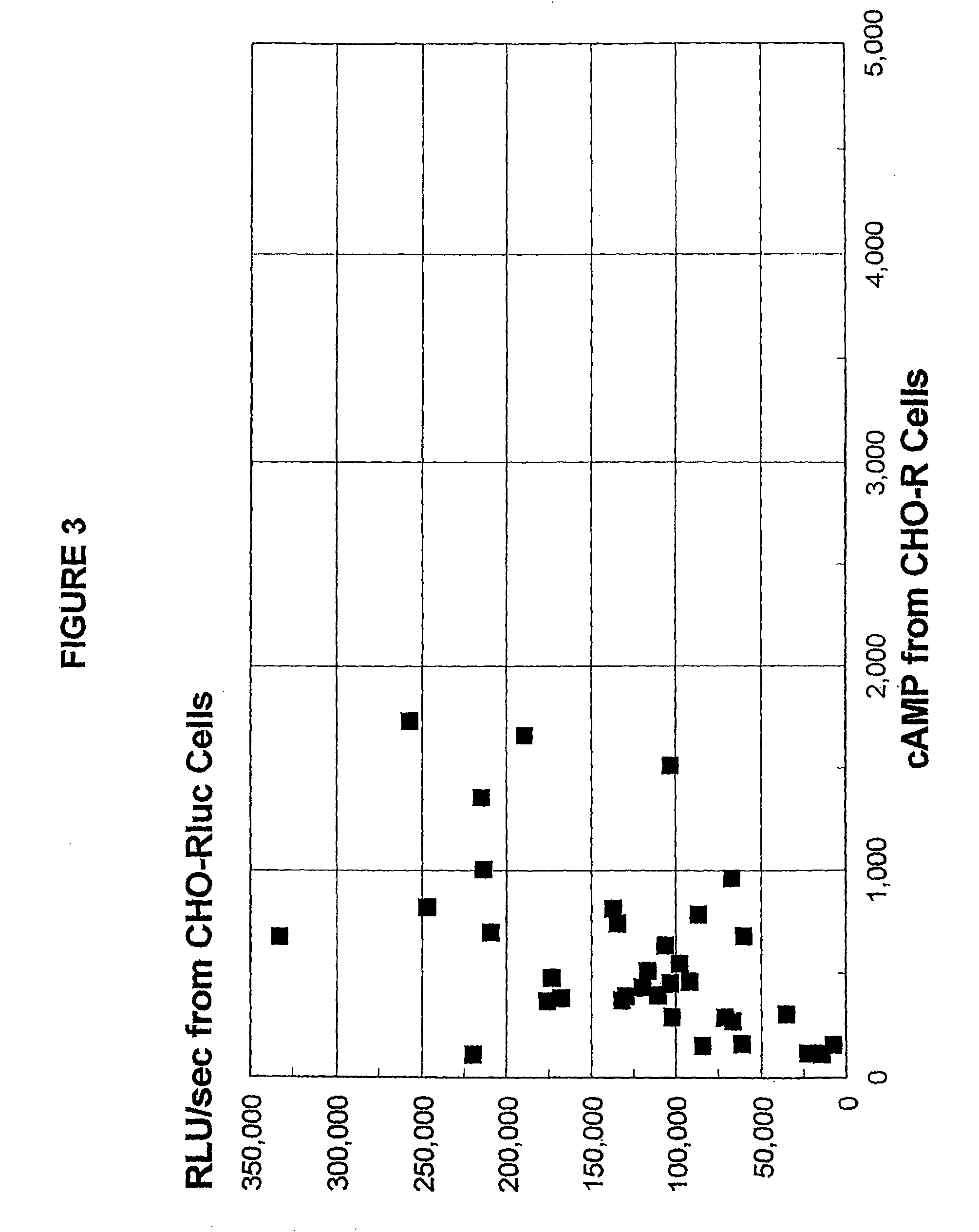
[0217]In these experiments, patients' IgG was prepared for testing in the present methods. Lyophilized IgG samples from 38 well-known and characterized, untreated Graves' disease patients were kindly provided by Dr. B. Y. Cho (Department of Internal Medicine, Seoul National University, College of Medicine, Seoul, Korea). As most of the samples had been previously tested in standard methods using CHO-R and FRTL-5 cells, these test results were known for 35 of these samples.

[0218]In preparation for lyophilization, the IgGs were affinity-purified using protein A-Sepharose CL-4B columns, as known in the art, and then dialyzed against 100 volumes of distilled water at 4° C. The dialysis water was changed every 8 hours over a 2 day period. After removal of denatured protein by centrifugation at 1500×g for 15 minutes at 4° C., the IgG was lyophilized and stored at −20° C. until used in the experiments described herein.

[0219]In some experiments, purified untreated ...


PUM


Abstract

The present invention provides methods and compositions useful in the diagnosis and management of autoimmune diseases. In particular, the present invention provides improved methods and compositions for the diagnosis and management of Graves' disease. The methods of the present invention not only avoids the need for radioactivity and are much simpler, economical, and rapid than methods traditionally used for the diagnosis of Graves' disease, but also improve upon the sensitivity and detection abilities of previous luciferase-based autoantibody detection assays. Such improvements are based upon the superior performance of assays comprising a chimeric TSH receptor in the presence of a glucocorticoid including, but not limited to, dexamethasone.

Description

STATEMENT OF RELATED APPLICATIONS[0001]This is a Continuation-In-Part of co-pending application(s) PCT / US2008 / 011027 with an International Filing Date of Sep. 24, 2008 which is a Continuation-In-Part of U.S. patent application Ser. No. 11 / 906,189 (now issued as U.S. Pat. No. 8,293,879) filed on Oct. 1, 2007 which is a Continuation-In-Part of U.S. patent application Ser. No. 10 / 996,961 filed on Nov. 24, 2004 (now abandoned) which is a divisional of U.S. patent application Ser. No. 09 / 539,735 (now issued as U.S. Pat. No. 6,852,546) filed on Mar. 30, 2000.FIELD OF THE INVENTION[0002]The present invention provides methods and compositions useful in the diagnosis of autoimmune diseases. In particular, the present invention provides methods and compositions for use in the diagnosis and management of Graves' disease. For example, one composition comprises a chimeric thyroid stimulating hormone receptor having improved sensitivity and specificity for circulating thyroid stimulating immunogl...

Claims


Application Information

Patent Timeline
Patent Type & Authority Patents(United States)
IPC IPC(8): G01N33/53
CPCC07K14/72G01N33/566C12Q1/6883C12N5/0602
Inventor KOHN, LEONARDBROWN, JIMSCHOLL, DAVIDLI, YUNSHENGNAPOLITANO, GIORGIO
Owner DIAGNOSTIC HYBRIDS
Why Eureka
Try Eureka