Method and apparatus for treatment of amblyopia

a technology for amblyopia and amblyopia, applied in the field of amblyopia apparatus and methods, can solve the problems of inability to develop additional synapses, inability to provide synapses, and abnormal outcomes, and achieve the effects of reducing or suspending impulse transmission, and facilitating catheter entry into the orbi

Inactive Publication Date: 2004-02-17
GUERRERO JOHN M
View PDF4 Cites 50 Cited by
  • Summary
  • Abstract
  • Description
  • Claims
  • Application Information

AI Technical Summary

Benefits of technology

The safety of 1% preservative-free lidocaine in the anterior segment of the eye is well established as the drug is frequently used as an anesthetic adjunct to topical anesthesia in modern cataract surgery. Anecdotal reports of 1% preservative-free lidocaine entering the posterior segment of the eye (Hoffman and Fine, 1997) have demonstrated that the drug blocked transmission of visual signals to the patient's brain as the patient rapidly developed "no light perception" vision. This effect was reversible, however, and vision returned in the eye within hours of discontinuing the drug. The authors report no evidence of retinal toxicity following the exposure to 1% preservative-free lidocaine.
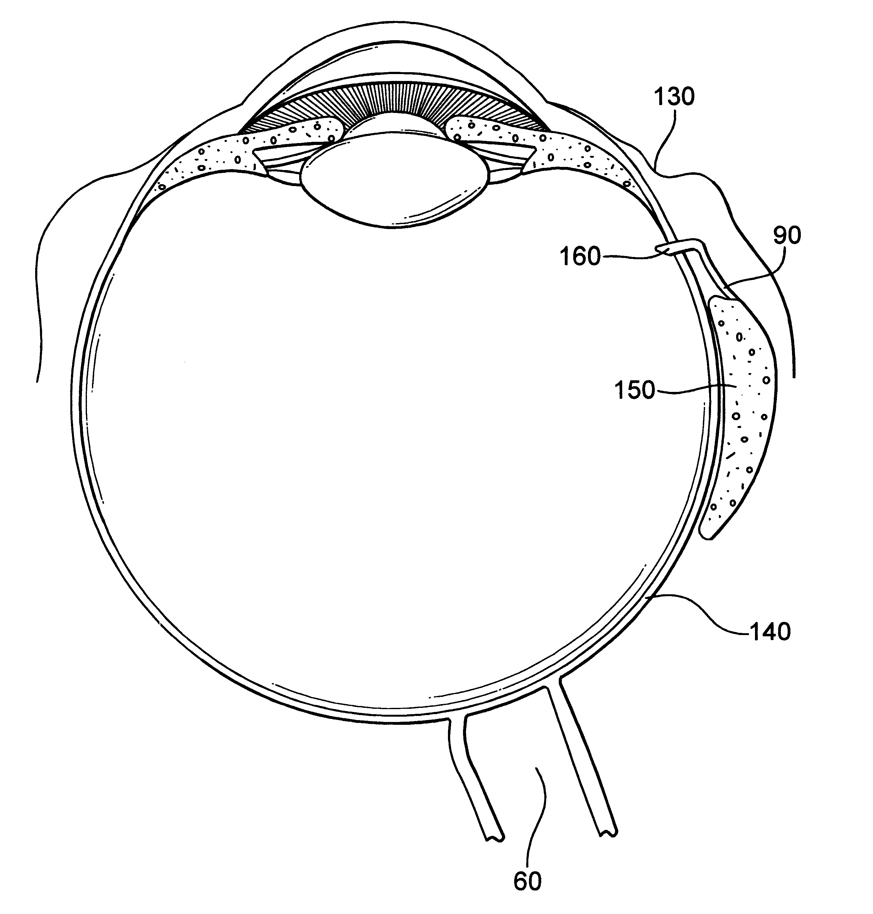
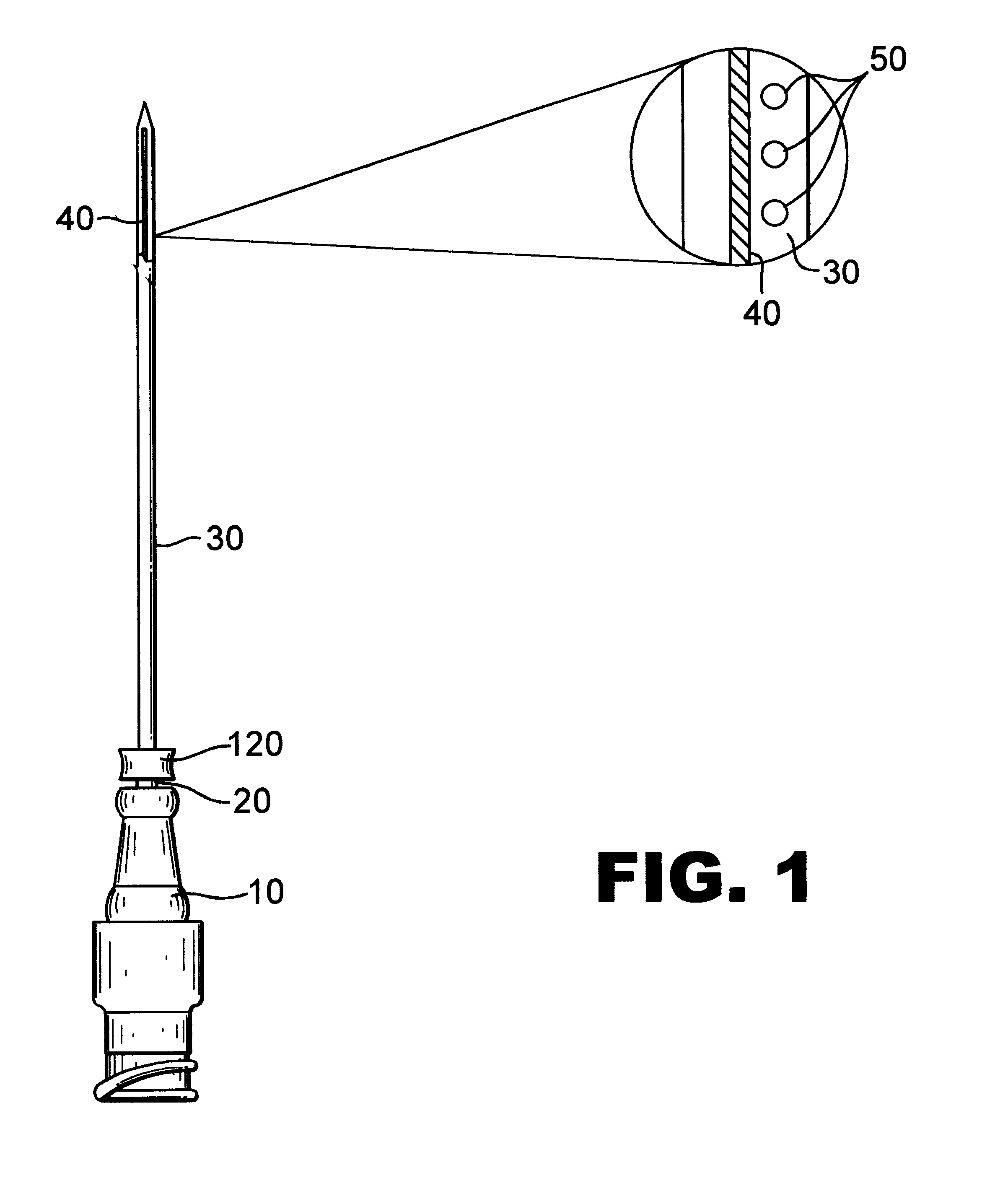
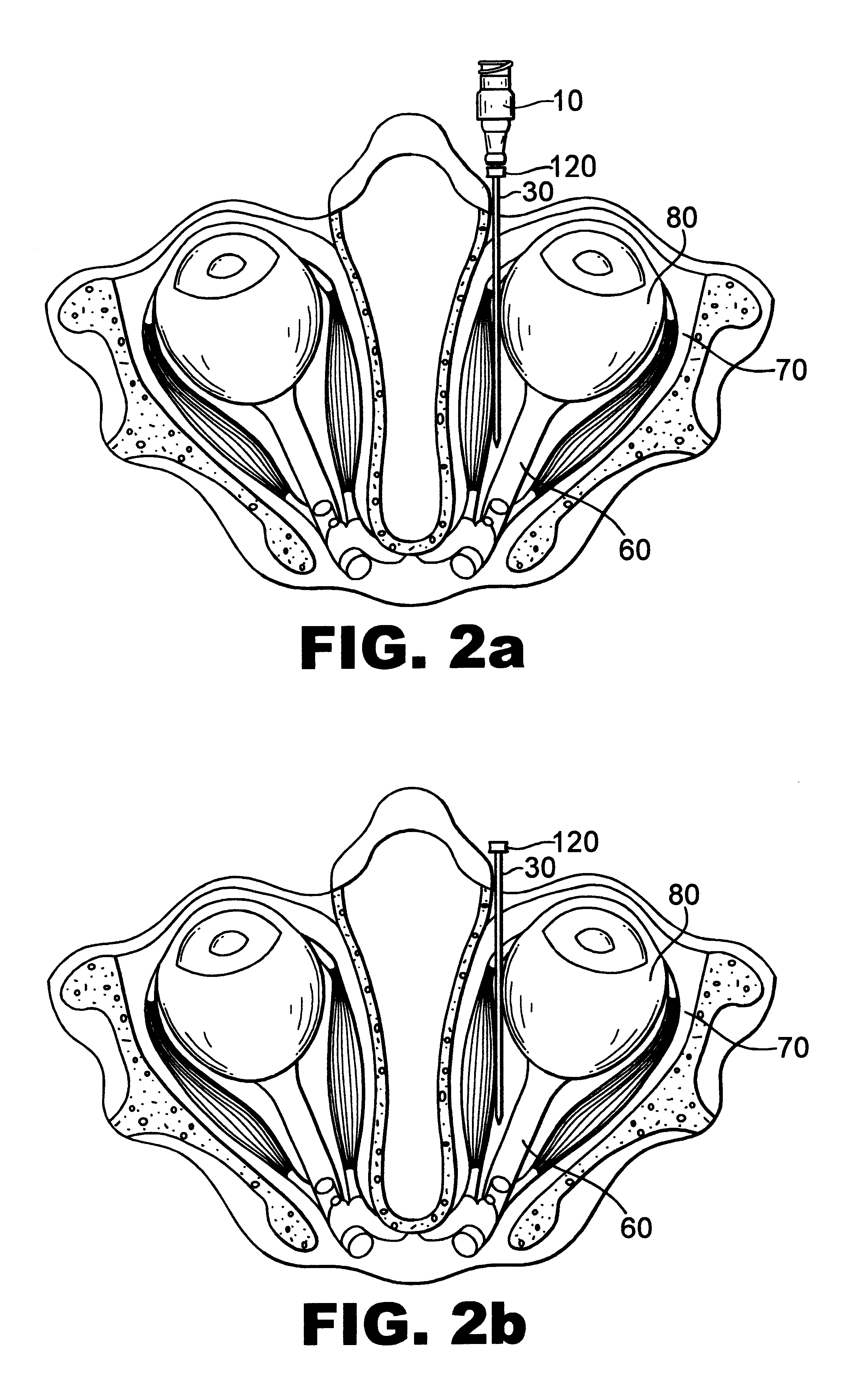
Another aspect of the invention is a method of providing controlled amounts of the selected drug to the targeted nerve. In one embodiment, a device employing a small drug delivery pump directs an appropriate drug through a canula into the orbit (socket) of the dominant eye. A fenestrated canula or catheter, preferably made of flexible, biocompatible material, is placed into the orbit of the dominant eye using one of several maneuvers. The catheter can be inserted without guidance into the orbit over a needle. A similar "blind stick" technique is used to induce peribulbar or retrobulbar anesthesia prior to ocular surgery. The needle is then withdrawn, leaving the catheter in the orbit. The catheter can also be advanced into the orbit under CT (computerized tomography) or fluoroscopic guidance. Finally, guidance of the catheter into the orbit may be facilitated by electronic feedback. The infusion catheter or canula can include a conductive strip allowing the catheter to function as a sensing electrode. Alternatively, the infusion catheter can be introduced over a needle possessing an insulated shaft and a conductive tip such that the needle acts as a sensing electrode. In either case, photic stimulation is provided to the dominant eye thereby producing impulses in its optic nerve which are detected by the electrode as it comes in closer and closer proximity to the optic nerve. In this way, feedback from the electrode allows the surgeon to accurately place the catheter near the optic nerve while feedback from the electrode allows the surgeon to avoid traumatizing the optic nerve with the needle or catheter.
The catheter may also be placed in close proximity to the optic nerve by direct visualization using a surgical approach such as a medial orbitotomy. While more invasive, this method would allow exact placement of the canula near the optic nerve. Using this technique, placement of the canula could be accompanied by optic nerve sheath fenestration, a technique in which a "window" is made into the dural covering of the optic nerve, thereby allowing any infused drugs easier access to the optic nerve.
Following catheter placement, a drug delivery pump supplies an infusion of drug sufficient to decrease or suspend impulse transmission by the optic nerve. The pump is easily miniaturized so that it can be comfortably placed on the neck or behind the ear of the patient or even implanted beneath the conjunctiva with little discomfort or inconvenience. Control of drug delivery can be by manual set in the physician's office, or adjusted automatically through use of a microprocessor. The microprocessor can be adapted to detect signals from the dominant eye to the optic nerve so that an amount of drug can be administered that is effective to prevent or inhibit the transmission of impulses from the dominant eye to the brain via the optic nerve.

Problems solved by technology

This is thought to result from an abnormal outcome of the competitive process of visual development.
The number of synapses available within a given area of cortex is limited and once this number of synapses is reached, the development of additional synapses cannot occur without the loss or destruction of other synapses in the same cortical area.
In amblyopia, one eye is disadvantaged relative to the contralateral eye.
Second, the better-seeing eye is temporarily disadvantaged relative to the amblyopic eye.
In preverbal children, the task is significantly more difficult and often involves the examiner observing the child's ability to fixate upon and follow a small target with each eye.
Occlusive therapy is problematic for several reasons: First, the technique is only effective during the so-called critical period during which the visual system is developing.
The older the patient is, the less successful occlusive therapy tends to be, with occlusive therapy in adults having little or no significant value.
Second, and of significant importance, keeping an occlusive dressing over the dominant eye of a young child is a challenge for child, parent, and physician alike.
Understandably, many children fail occlusive therapy due to noncompliance.
This leaves these patients with permanent visual loss in the amblyopic eye, as occlusion therapy is not efficacious in adults and there are currently no effective methods for the treatment of adult amblyopia.
The challenge in an amblyopic patient therefore is to improve vision in the amblyopic eye without causing significant reduction of visual acuity in the dominant eye.
There is currently no such therapy available to adult patients or to pediatric patients refractory to occlusion therapy.
The lack of a demonstrably effective treatment for adult amblyopia leaves many patients with severely limited, often debilitating, vision.

Method used


Image

  • Method and apparatus for treatment of amblyopia
  • Method and apparatus for treatment of amblyopia
  • Method and apparatus for treatment of amblyopia

Examples


example 1

5.1 Example 1

Trauma to Dominant Eye

A 33 year-old male has a history of dense (20 / 200) amblyopia in the right eye. He was 20 / 20 in his dominant left eye prior to a motor vehicle accident in which he sustained blunt trauma with extensive choroidal hemorrhage to his left eye. One year following his injury, he has 20 / 200 vision in his amblyopic right eye and 20 / 400 in his previously dominant left eye. The patient wishes to improve vision in his amblyopic right eye to permit reading, allow him to return to work, and possibly allow him to drive. A drug delivery canula is placed through the pars plana of the formerly dominant left eye using a standard trans-pars plana vitrectomy technique. This canula is connected to a small drug delivery pump, which is then implanted beneath the conjunctiva of left eye. A nerve-blocking agent in this case 1% preservative-free lidocaine, is delivered to the eye in sufficient quantity to block transmission of impulses from the dominant left eye to the brain...

example 2

5.2 Example 2

Corneal Scarring in Dominant Eye

A 43 year-old female with a history of amblyopia in her right eye (20 / 100) develops HSV (Herpes Simplex Virus) keratitis in her dominant (20 / 20) left eye. Dense corneal scars result in 20 / 200 vision in her dominant left eye. Multiple corneal transplants are attempted in the patient's dominant eye and fail due to recurrent HSV. The patient is left with 20 / 100 vision in her amblyopic eye and "count fingers" vision in her right eye. Likewise, this patient wishes to improve the vision in her amblyopic eye, preferably to levels compatible with safely driving an automobile. In the operating room, a flexible catheter is positioned in the orbit (socket) of the patient's dominant left eye adjacent to the optic nerve. This catheter is then connected via a segment of tubing to a drug delivery pump that is then sutured to the sclera and implanted beneath the conjunctiva of the eye. The pump delivers a reversible denervating drug, in this case 0.50% b...

example 3

5.3 Example 3

Penetrating Trauma to a Dominant Eye

A 13 year-old male with a history of amblyopia in his left eye (20 / 200 visual acuity) was using a lawn mower when it ejected a stone, which struck and penetrated his dominant right eye (20 / 15 visual acuity). Following surgical repair, he is left with "light perception only" vision in his dominant right eye. The patient desires to improve the vision in his amblyopic eye so as to resume normal activities of daily living, including attending school. As with most patients who sustain severe ocular trauma, this patient requires multiple surgical interventions to save the eye and what little visual acuity remains. During a retinal detachment repair of the traumatized dominant right eye, an intraocular drug delivery insert is implanted in the eye using standard surgical techniques. The insert delivers a reversible denervating agent such as 1% preservative-free lidocaine to the dominant eye, allowing recovery of vision in the amblyopic eye. T...


PUM


Abstract

A method of improving vision in an amblyopic eye is described in which vision in the dominant eye is reversibly disrupted. The technique promotes improvement of visual acuity in the weaker eye. The disclosed method is particularly suitable for treatment of adult amblyopia. The method also includes several devices suitable for use in the described treatments.

Description

1.0 BACKGROUND ART1.1 Field of the InventionThe invention concerns apparatus and methods for treating amblyopic conditions and is particularly directed to methods that correct this visual deficiency in adults.1.2 Description of Related ArtAmblyopia, Greek for "blunt vision", is the failure of an anatomically intact eye to develop normal visual acuity. Amblyopia affects five to seven million individuals in the United States alone, with an estimated 70,000 new cases annually. Amblyopia accounts for more visual loss in the under-45 age group than all other ocular diseases, including trauma. In the lay press, this condition is often referred to as "lazy eye". The term lazy eye is to be avoided, however, as it may also be used in the lay press to describe strabismus ("crossed" eye) as well. While a crossed eye may become amblyopic (strabismic amblyopia), not all crossed eyes are amblyopic nor are all amblyopic eyes crossed.Amblyopia represents a failure of the affected eye to develop nor...

Claims


Application Information

Patent Timeline
Patent Type & Authority Patents(United States)
IPC IPC(8): A61F9/00A61M31/00A61M25/06
CPCA61F9/0017A61M31/00A61M25/0606
Inventor GUERRERO, JOHN M.
Owner GUERRERO JOHN M
Why Eureka
Try Eureka