
Magnetic resonance imaging method and apparatus employing partial parallel acquisition, wherein each coil produces a complete k-space datasheet

Inactive Publication Date: 2005-01-11
GRISWOLD MARK
View PDF10 Cites 157 Cited by
  • Summary
  • Abstract
  • Description
  • Claims
  • Application Information

AI Technical Summary

Benefits of technology

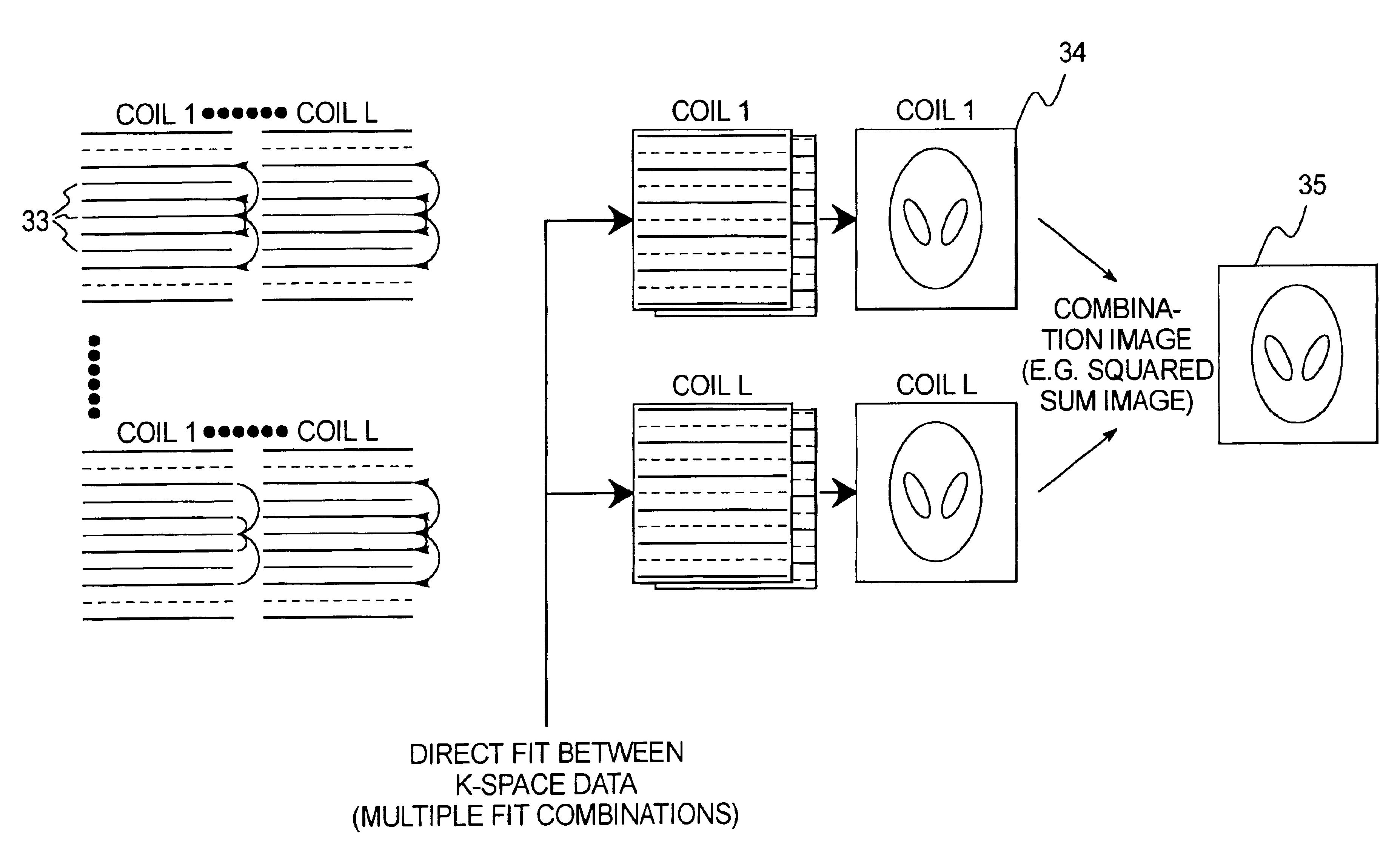
It is an object of the present invention to provide an apparatus and a method for magnetic resonance imaging that enable a better quality of the reconstructed images on the basis of a partially parallel acquisition (PPA).

Problems solved by technology

Despite the technical progress in the construction of MRI apparatuses, the exposure time of an MRI image remains the limiting factor for many applications of MRI in medical diagnostics.
Further enhancement in the performance of MRI apparatus is limited from a technical point of view (feasibility) and for reasons of patient protection (stimulation and tissue heating).
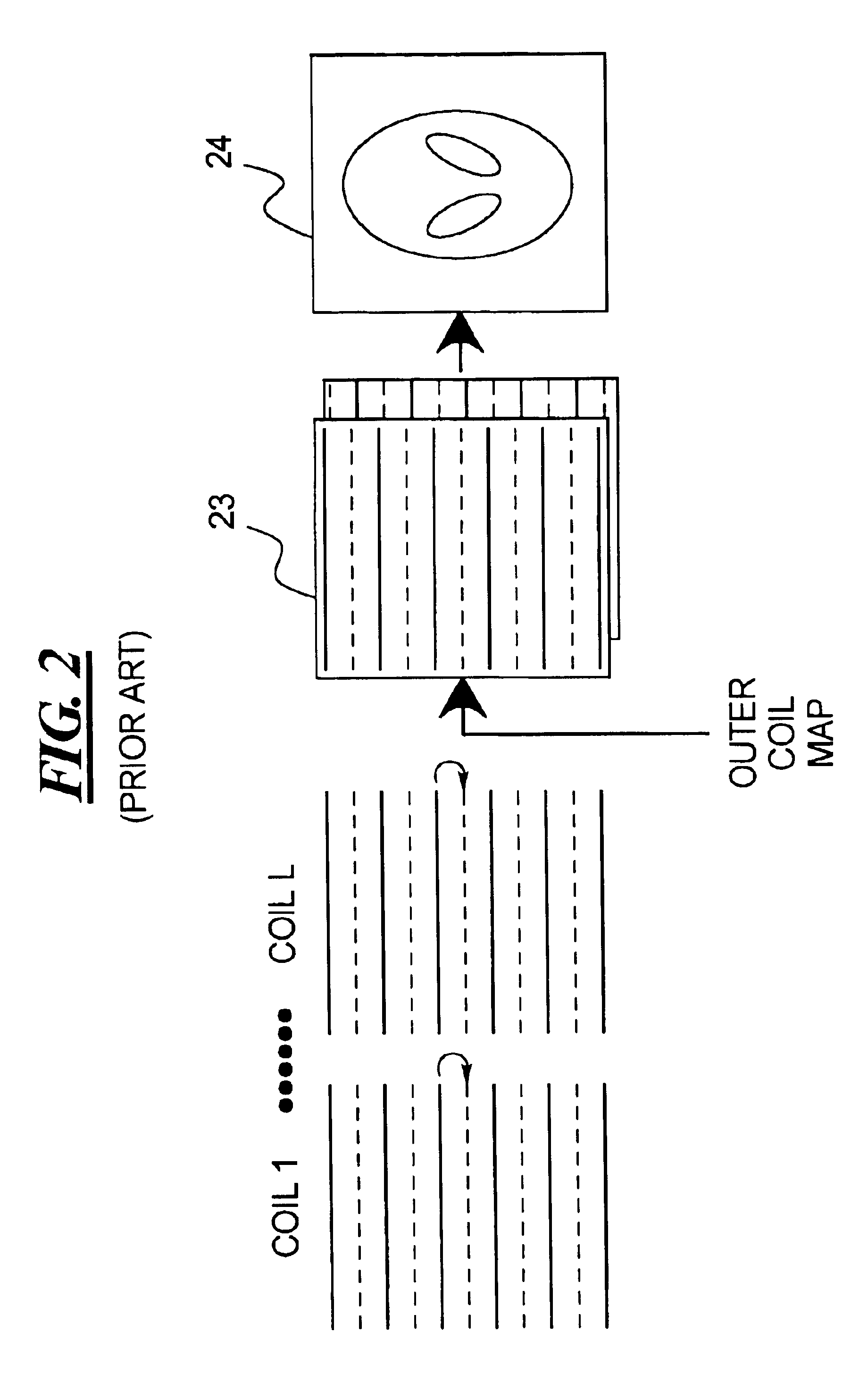
Whereas some of these PPA techniques (SMASH, SENSE, GSMASH, which are described in brief below) have been successfully employed in many areas of MRI —SMASH and SENSE must be given priority—, the most significant disadvantage of these methods is that the complex sensitivity of every individual component coil must be exactly known.
This is frequently problematical in practice since the experimental determination of the coil sensitivities is greatly hindered due to disturbances as a result of noise and—even more important—due to spin density fluctuations in the tissue, and thus leads to error-affected reconstructions.
It is specifically this problem that still restricts a widespread clinical application of PPA methods.
Due to disturbances as a result of noise and spin density fluctuations within the subject, it is usually extremely difficult to determine this.
Whereas it has been shown that the AUTO-SMASH method functions well in some instances, it has been shown in many other instances how noise and weak coil signal degrade the AUTO-SMASH reconstruction, particularly given high acceleration factors.
As a result, reconstructed and, consequently, error-affected lines are replaced.
Moreover, these lines can be employed in the final image reconstruction, which leads to a lower artifact energy.
A principal problem of all SMASH-like reconstructions—in addition to the termination of coil sensitivities—is that the S / N of the reconstructed image corresponds only to that of a phase-summed image, since there is no possibility of forming a square sum image.
In addition to the inherent S / N losses, errors also arise because the k-space data of the various coils in a SMASH-typical reconstruction are combined by complex addition.
This method of determining the appropriate coil phases can fail in many instances, which may lead to an unsatisfactory image quality and / or to serious phase cancellation.
The reconstruction of all known PPA techniques ensues by complex addition of the individual picture elements, which can lead to reconstruction artifacts (phase cancellation of anti-phase elements).

Method used


Image

  • Magnetic resonance imaging method and apparatus employing partial parallel acquisition, wherein each coil produces a complete k-space datasheet
  • Magnetic resonance imaging method and apparatus employing partial parallel acquisition, wherein each coil produces a complete k-space datasheet
  • Magnetic resonance imaging method and apparatus employing partial parallel acquisition, wherein each coil produces a complete k-space datasheet

Examples


Embodiment Construction

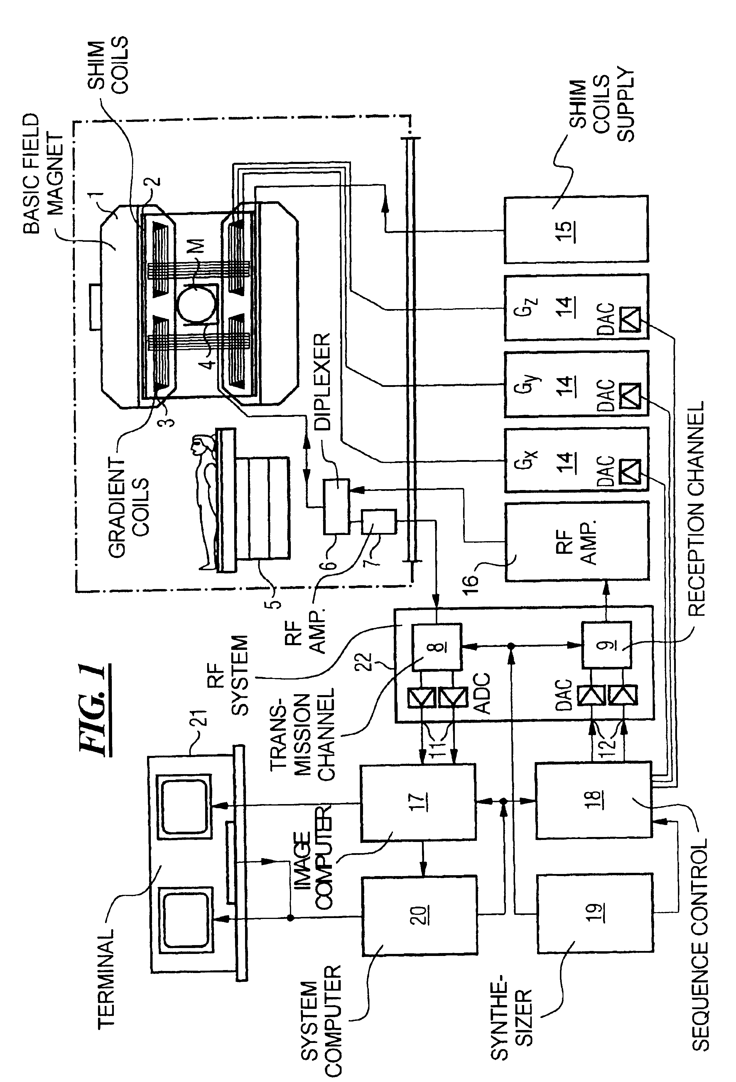
FIG. 1 schematically illustrates a magnetic resonance imaging (tomography) apparatus for generating a nuclear magnetic image of a subject according to the present invention. The components of the nuclear magnetic resonance tomography apparatus correspond to those of a conventional tomography apparatus, but it is controlled according to the invention. A basic field magnet 1 generates a time-constant, intense magnetic field for polarization (alignment) of the nuclear spins in the examination region of a subject such as, for example, a part of a human body to be examined. The high homogeneity of the basic magnetic field required for the nuclear magnetic resonance measurement is defined in a spherical measurement volume M in which the part of the human body to be examined is introduced. For supporting the homogeneity demands and, in particular, for eliminating time-invariable influences, shim plates of ferromagnetic material are attached at suitable locations. Time-variable influences a...


PUM


Abstract

In a method and apparatus for magnetic resonance imaging of an interconnected region of a human body on the basis of a partially parallel acquisition (PPA) by excitation of nuclear spins and measurement of the radio-frequency signals produced by the excited nuclear spins, a number of spin excitations and measurements of an RF response signal are implemented simultaneously in every component coil of a number of RF reception coils. As a result a number of response signals are acquired that form a reduced dataset of received RF signals for each component coil. Additional calibration data points are acquired for each reduced dataset. A complete image dataset is formed for each component coil on the basis of the reduced dataset for that component coil and at least one further, reduced dataset of a different component coil. A spatial transformation of the image dataset of each component coil is implemented in order to form a complete image of each component coil.

Description

BACKGROUND OF THE INVENTION1. Field of the InventionThe present invention is directed in general to nuclear magnetic resonance tomography (also called magnetic resonance imaging—MRI) as employed in medicine for examining patients. The present invention is particularly directed to a magnetic resonance tomography apparatus as well as to a method for the operation thereof wherein the technique referred to as partially parallel acquisition (PPA) is employed.2. Description of the Prior ArtMRI is based on the physical phenomenon of nuclear magnetic resonance and has been successfully utilized as an imaging method in medicine and in biophysics for more then fifteen years. Given this examination method, the subject is subjected to a strong, constant magnetic field. As a result, the nuclear spins of the atoms in the subject, that were previously irregularly oriented, are aligned. Radio-frequency pulses can then excite these “ordered” nuclear spins to a specific resonance. This resonance gene...

Claims


Application Information

Patent Timeline
IPC IPC(8): G01R33/561G01R33/54
CPCG01R33/5611
Inventor GRISWOLD, MARK
Owner GRISWOLD MARK
Learn More