Method and apparatus for calibrating detector spectral response

a detector and spectral response technology, applied in the field of medical imaging, can solve the problems of uniformity in the reconstructed image of a uniform object, the reconstructed image may still contain ring or band artifacts, and achieve the effect of rapid calibration of the spectral response and greater coverage of the detector elemen

Inactive Publication Date: 2005-02-01
GENERAL ELECTRIC CO
View PDF3 Cites 17 Cited by
  • Summary
  • Abstract
  • Description
  • Claims
  • Application Information

AI Technical Summary

Benefits of technology

The present technique provides a novel approach for the spectral calibration of the detector elements of a CT detector using one or more calibration phantoms offset from isocenter. The offset phantoms provide greater coverage of the detector elements and provide spectral response data for a range of penetration lengths for each detector element. The penetration lengths range in length from a minimum chord of the phantom, determined by the offset distance and the diameter of the phantom, to the diameter of the phantom. For each detector element, the spectral response as a function of projection value can be obtained by comparing the fitting of each projection view to the corresponding measured projection view over all view angles. The fitting information may then be employed to derive the coefficients of the spectral response curve for each detector element, which may in turn be employed to provide rapid calibration of the spectral response for each element.

Problems solved by technology

In particular, the beam-hardening phenomena may cause nonuniformities in a reconstructed image of a uniform object, such as the phantoms used in calibration.
With such a beam hardening spectral correction, the resulting reconstructed image, however, may still contain ring or band artifacts due to the differential spectral response of the various detector elements.
Various factors, however, may result in the derivation of correction functions from the spectral calibration process that are insufficient or inadequate to fully remove artifacts in the reconstructed image resulting from differential detector element response.
In particular, the techniques employed to derive correction functions typically rely upon an insufficient number of data points representing the spectral response of an element as a function of projection value.
As a result, calibration data are acquired at each detector element for a single penetration length for each phantom and do not provide information about detector spectral response as a function of X-ray penetration length.
The resulting correction function, however, is generally substantially linear and may fail to adequately correct the differential spectral responses of the detector elements to the extent that such responses are non-linear away from the measured data points.
In addition, the image regions corresponding to the joining of the different data sets may give rise to image artifacts.
This technique, however, may be unreliable if the detector elements differentially introduce large relative spectral errors, which influence the computation of the smoothed projections, resulting in the extraction of an incorrect baseline.

Method used


Image

  • Method and apparatus for calibrating detector spectral response
  • Method and apparatus for calibrating detector spectral response
  • Method and apparatus for calibrating detector spectral response

Examples


Embodiment Construction

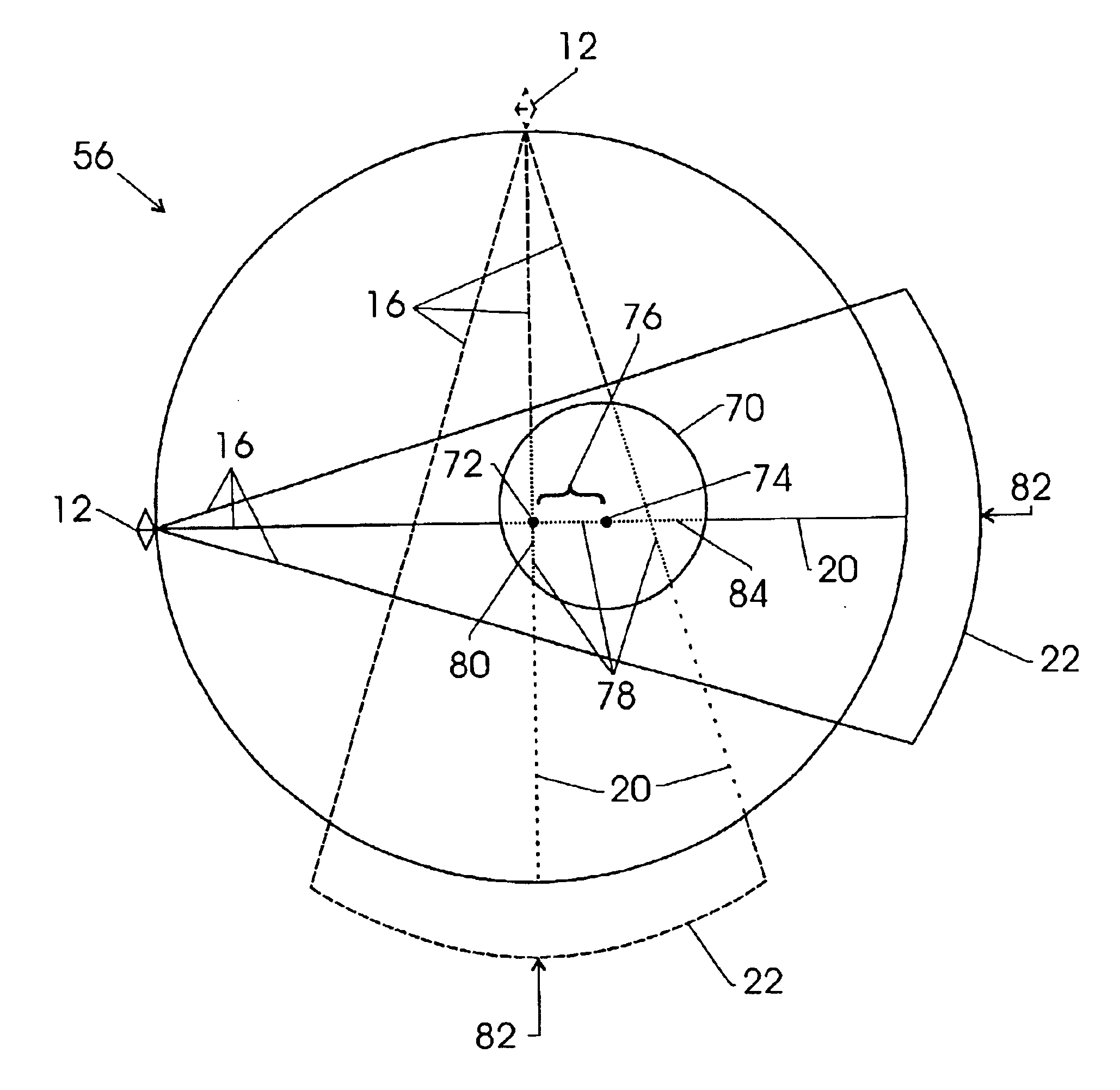
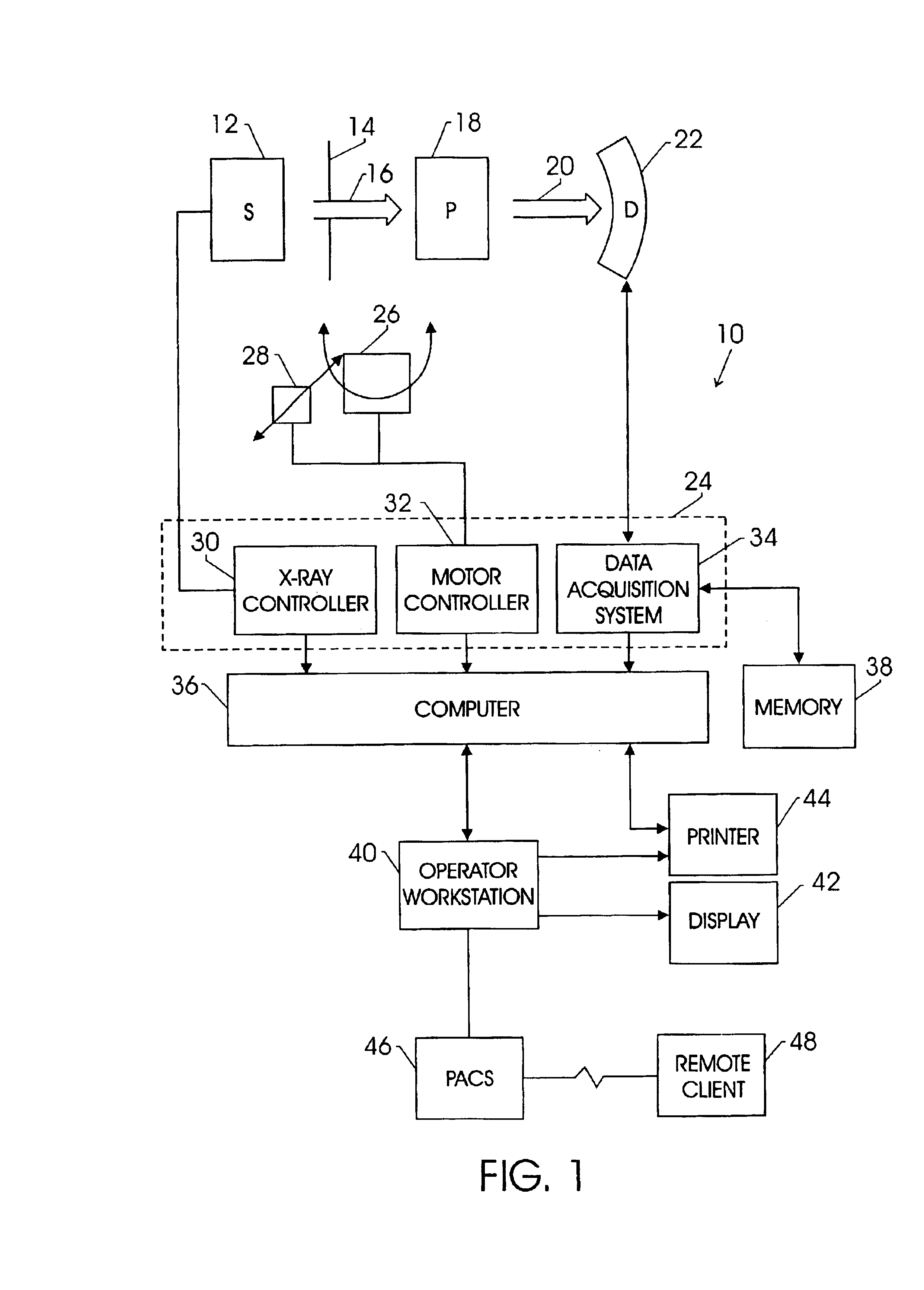
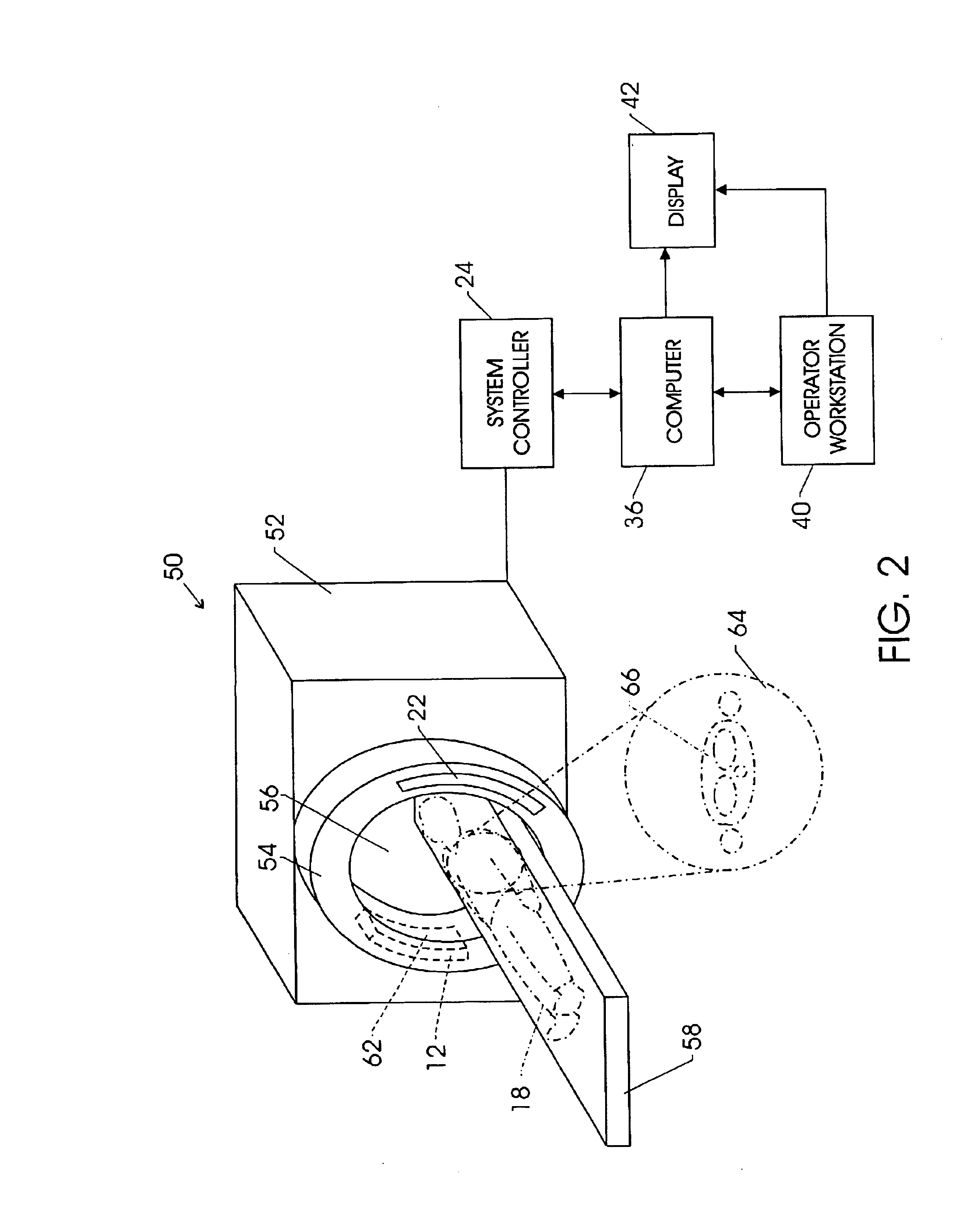
FIG. 1 illustrates diagrammatically an imaging system 10 for acquiring and processing image data. In the illustrated embodiment, system 10 is a computed tomography (CT) system designed both to acquire original image data, and to process the image data for display and analysis in accordance with the present technique. In the embodiment illustrated in FIG. 1, imaging system 10 includes a source of X-ray radiation 12 positioned adjacent to a collimator 14. In this exemplary embodiment, the source of X-ray radiation source 12 is typically an X-ray tube.

Collimator 14 permits a stream of radiation 16 to pass into a region in which a subject, such as a human patient 18 is positioned. A portion of the radiation 20 passes through or around the subject and impacts a detector array, represented generally at reference numeral 22. Detector elements of the array produce electrical signals that represent the intensity of the incident X-ray beam. These signals are acquired and processed to reconstr...


PUM


Abstract

The present technique provides for the spectral calibration of the detector elements of a CT detector using one or more offset calibration phantoms. The offset phantoms provide greater coverage of the detector elements as well as spectral response data associated with penetration lengths ranging in length from a minimum chord of the phantom to the diameter of the phantom. The spectral response as a function of penetration length can be obtained for each detector element by comparing the fitting of each projection view to the corresponding measured projection view over all view angles. The fitting information may then be employed to derive the coefficients of the spectral response curve for each detector element, which may in turn be employed to provide rapid correction of the spectral response for each element.

Description

BACKGROUND OF THE INVENTIONThe present invention relates generally to the field of medical imaging. In particular, the following techniques relate to computed tomography imaging systems and the calibration of detectors used in such systems.Computed tomography (CT) imaging systems measure the attenuation of X-ray beams passed through a patient from numerous angles. Based upon these measurements, a computer is able to reconstruct images of the portions of a patient's body responsible for the radiation attenuation. As will be appreciated by those skilled in the art, these images are based upon separate examination of a series of angularly displaced projection images. A CT system produces data that represents the line integral of linear attenuation coefficients of the scanned object. This data is then reconstructed to produce an image, which is typically displayed on a cathode ray tube, and may be printed or reproduced on film. A virtual 3-D image may also be produced by a CT examinatio...

Claims


Application Information

Patent Timeline
Patent Type & Authority Patents(United States)
IPC IPC(8): A61B6/00
CPCA61B6/583
Inventor WU, XIAOYELEBLANC, JAMESWALTER, DEBORAH J.
Owner GENERAL ELECTRIC CO
Why Eureka
Try Eureka