

Method for the detection, identification, enumeration and confirmation of virally infected cells and other epitopically defined cells in whole blood
a technology of epitopically defined cells and whole blood, applied in the field of whole blood whole blood enumeration, detection, identification, enumeration and confirmation of virally or nonvirally infected cells, can solve the problems of serology not providing quantitative information, high cost, and inconvenient assembly, so as to reduce the possibility of false positive results
- Summary
- Abstract
- Description
- Claims
- Application Information
AI Technical Summary
Benefits of technology
Problems solved by technology
Method used
Image

Examples
Embodiment Construction
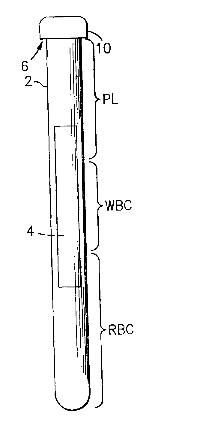
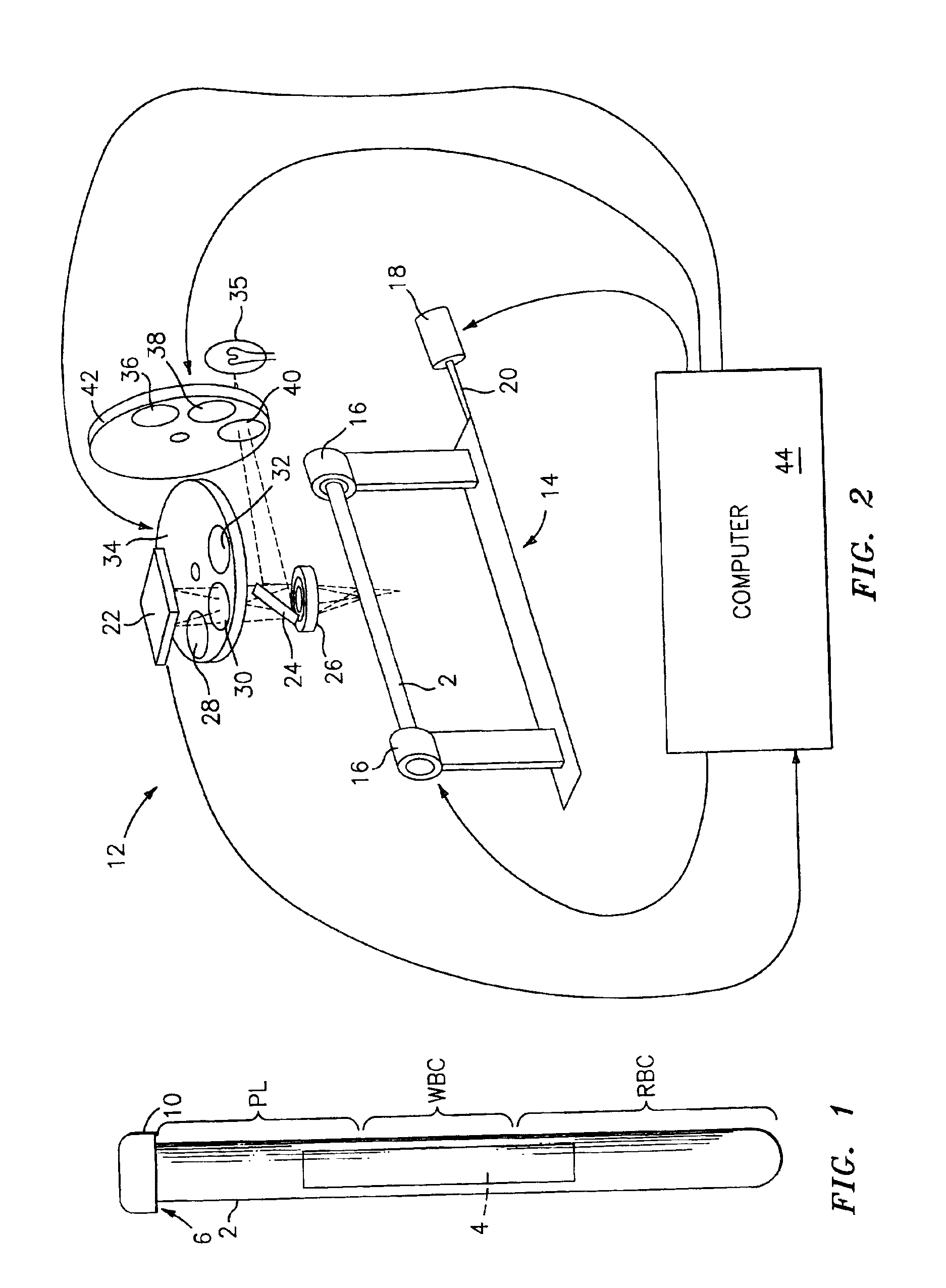
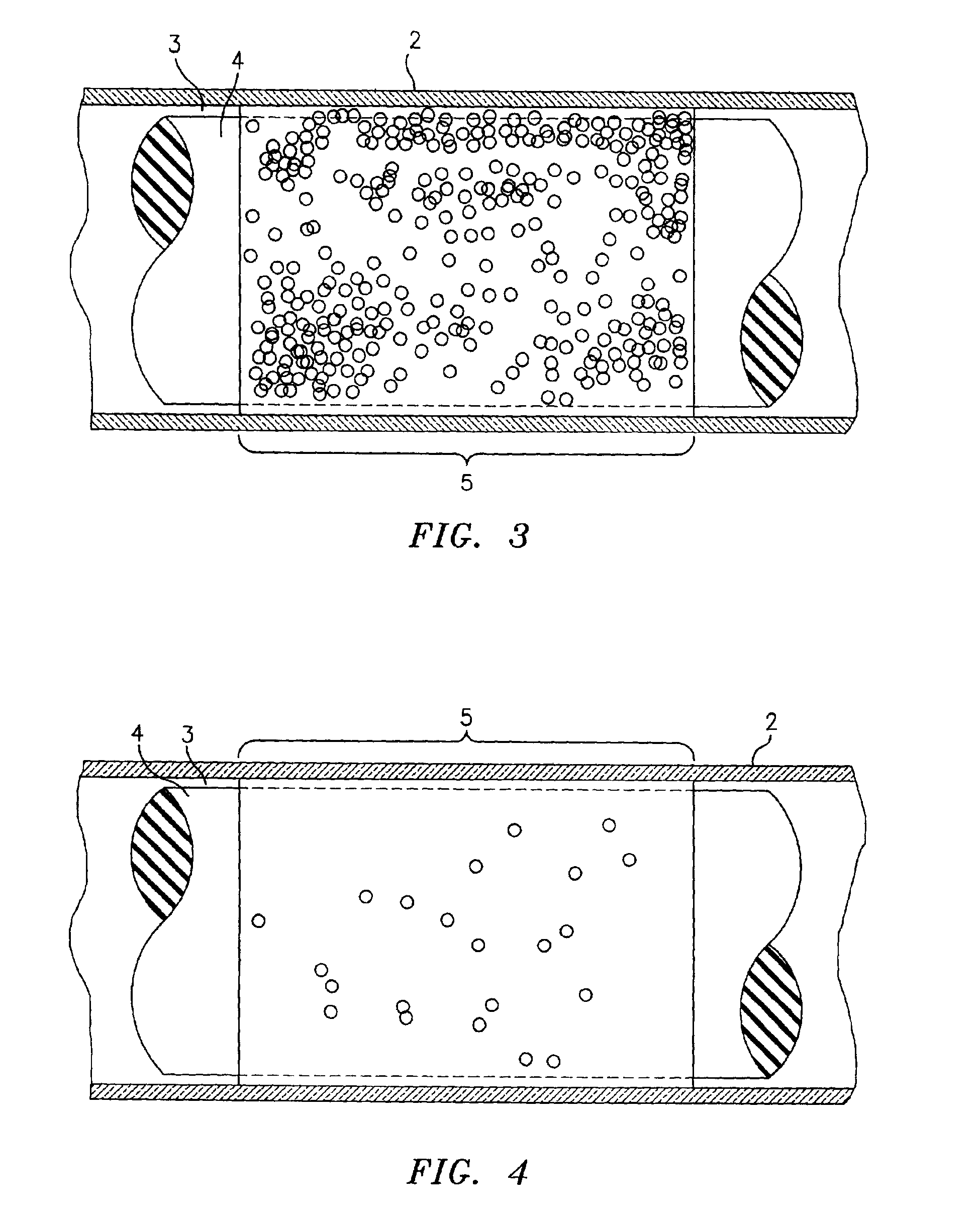
[0022]Referring now to the drawings, there is shown in FIG. 1 a side elevational view of a sampling tube and and insert assembly, which is referred to hereinafter generally as “the paraphernalia” and which includes a transparent blood sampling tube 2 that contains an elongated plastic insert 4. The tube 2 has an upper end 6 which is closed off by means of a closure cap 10. The tube 2 is a larger tube which is sized to contain about 10 mls of blood. The thickness of the gap between the tube bore and the insert 4 will be larger than the diameter of the target cells, and preferably will be equal to or less than the depth of the field of view of the detection instrument which is described in greater detail hereinbelow. The gap between the tube bore and the insert 4 will thus be accessible to target cells and the cells will be viewable in the gap by the detection instrument's optics. Generally, the red blood cells will be found in the section of the centrifuged blood sample labeled “RBC”...
PUM
| Property | Measurement | Unit |
|---|---|---|
| volume | aaaaa | aaaaa |
| volume | aaaaa | aaaaa |
| transparent | aaaaa | aaaaa |
Abstract
Description
Claims
Application Information