Cultured skin and method of manufacturing the same

a skin and culture technology, applied in the field of cultured skin, can solve the problems of many unsolved problems in the art, short cell division lifetime, and low differential plasticity capacity, and achieve the effect of high successful grafting ra

Inactive Publication Date: 2005-07-12
NIPRO CORP
View PDF13 Cites 97 Cited by
  • Summary
  • Abstract
  • Description
  • Claims
  • Application Information

AI Technical Summary

Benefits of technology

[0008]The present inventors have made dedicated efforts to provide a cultured reconstructive skin with a high successful grafting rate using cells collectable from tissues included in an umbilical cord originated from a human fetus, finally completing the present invention.

Problems solved by technology

However, epithelium cells of the tissues provided as origins of the above cells are those in which cell differentiation and aging have progressed, so that their cell division lifetime may be short and their capacity for differential plasticity may be small.
Furthermore, there are many unsolved problems in the art.
Ethical problems are caused in offering of an excess normal tissue obtained at the time of surgical operations, and so on.

Method used


Image

  • Cultured skin and method of manufacturing the same
  • Cultured skin and method of manufacturing the same
  • Cultured skin and method of manufacturing the same

Examples


example 1

Separation and Culture of Umbilical Epithelium Cells

[0074]An umbilical cord (30-60 cm in length, 9-15 mm in diameter) removed after birth was sufficiently washed three times with HBSS (Hanks' balanced salt solution) or PBS (Phosphate-Buffered Saline solution) to remove adherent blood. Then, both ends of the umbilical cord were tied with strings, respectively, followed by dipping the umbilical cord into a solution prepared by diluting a proteolytic enzyme “dispase”(manufactured by BECTON DICKINSON) with a medium (MCDB153-1 / 4 medium) having the following composition such that the concentration of the enzyme becomes 20% in the solution. In this case, the umbilical cord was hung by strings and was then dipped into the solution, while both ends (2 cm each) of the umbilical cord were not dipped in the solution, followed by leaving the umbilical cord as it is for 25 to 50 hours at 4° C.[0075]Medium: MCDB153-1 / 4 Medium (a medium prepared by mixing MCDB153 medium and DMEM (incl., 0-10% FBS) ...

example 2

Separation and Culture of Umbilical Myofibroblasts

[0081]In Example 1, after separating the umbilical epithelium cells, blood vessels were removed from the remaining umbilical tissue using a scalpel and a pair of tweezers. Then, the umbilical tissue after the removal of blood vessels was cut into pieces of about 5 mm square using a scalpel and a pair of tweezers. Small pieces were rested in the culture dish, while the DMEM medium (incl. 10% FCS) was poured into the culture dish such that one surface of the small piece was dipped in the medium.

[0082]After one week past, it was observed that umbilical myofibroblasts were generated around the tissue piece, followed by the addition of about 5 ml of the DMEM medium (incl. 10% FCS) such that the umbilical tissue was sufficiently submerged.

[0083]After that, the sufficient growth of umbilical myofibroblasts was allowed and then the umbilical tissue was removed. Consequently, umbilical myofibroblasts (about 106 cells) were collected using a s...

example 3

Proliferation of Separated and Cultured Umbilical Epithelium Cells

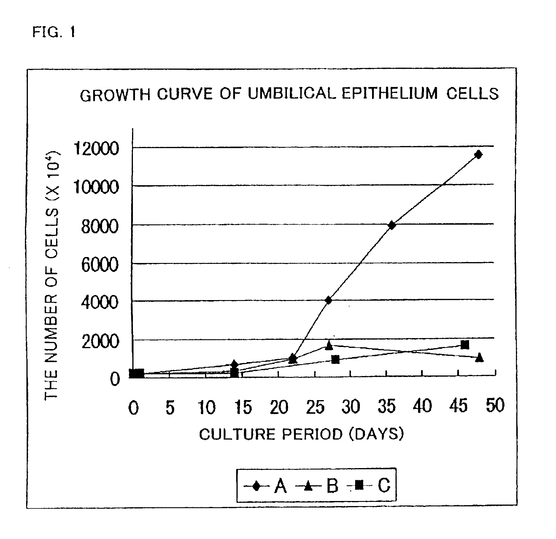
[0085]Umbilical epithelium cells separated in the same way as that of Example 1 were cultured using the following three kinds of media (A, B, C), respectively.

[0086]MCDB153-1 / 4(0% cx-FCS, 0% FCS) medium . . . (A)

[0087]Keratinocyte-SFM medium (GIBCO) . . . (B)

[0088]Defined Keratinocyte-SFM medium (GIBCO) . . . (C)

[0089]A subculture of the umbilical epithelium cells was prepared by collecting the cells using a solution of 0.1% trypsin and 0.1 mM EDTA in a state where the cells had became sub-confluent. The number of cells was counted, followed by inoculating the cells into a culture vessel with a volume three times higher than that of the prior vessel (the volume is the same as in Example 1).

[0090]The growth curve of the resultant cells is shown in FIG. 1.


PUM

PropertyMeasurementUnit
humidityaaaaaaaaaa
humidityaaaaaaaaaa
thicknessaaaaaaaaaa

Abstract

A cultured skin and a grafting cultured skin sheet are provided, each of which is a cultured reconstructive skin with a high take rate using cells collectable from cells originated from tissue included in an umbilical cord such as tissue included in an umbilical cord originated from a human fetus. The grafting cultured skin stratified sheet is prepared by placing an epithelium sheet on the top surface of a cultured dermis. The cultured dermis includes as components a cultured skin containing cells originated from a tissue included in an umbilical cord, such as umbilical cells, more concretely, umbilical fibroblast cells, being separated and cultured, preferably in a collagen nonwoven fabric. On the other hand, the epithelium sheet is prepared by culturing and stratifying the umbilical cord epithelium cells.

Description

TECHNICAL FIELD OF THE INVENTION[0001]The present invention relates to a cultured skin including cells originated from tissue included in an umbilical cord, such as umbilical cells, placental cells, and velamen cells. More specifically, the present invention relates to an artificial cultured skin to be used for skin grafting and cell therapy for various kinds of tissue reconstruction or regenerative medicine.BACKGROUND OF THE INVENTION[0002]A skin is provided as a boundary between a living body and an external environment and has functions of maintaining a liquid in the living body, reducing friction in water, keeping body temperature constant, and protecting the inside of the living body from physical damages.[0003]The skin of a vertebrate animal consists of a connective tissue layer generally regarded as dermis originated from the mesoderm and epidermis stratified thereon originated from the ectoderm.[0004]Cultured skin is constructed from a part of a skin structure by in vitro cu...

Claims


Application Information

Patent Timeline
Patent Type & Authority Patents(United States)
IPC IPC(8): A61L27/60A61L27/38A61L27/00C12N5/06A61F2/10C12N5/071
CPCA61L27/3804A61L27/3813A61L27/3826A61L27/383A61L27/3891A61L27/60C12N5/0698C12N2502/094C12N2502/1323C12N2506/02
Inventor YASUMOTO, SHIGERUTAKEUCHI, MASAKATSU
Owner NIPRO CORP
Why Eureka
Try Eureka